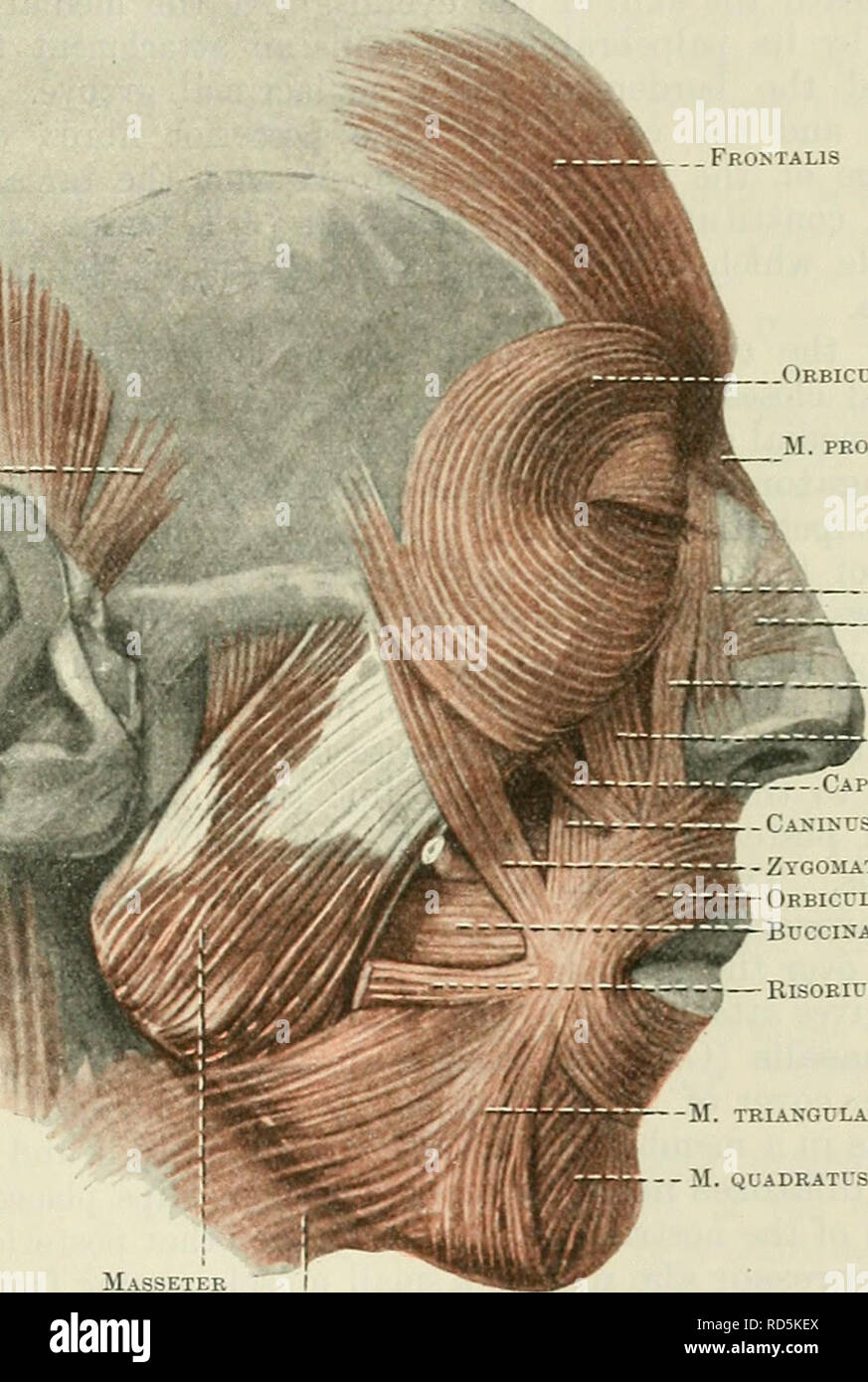
. Cunningham's Text-book of anatomy. Anatomy. THE MUSCLES OF THE SCALP. 449 The galea aponeurotica (O.T. epicranial aponeurosis), extending between the two fleshy bellies, is a continuous membrane which glides over the calvaria, and has attachments laterally to the temporal ridge, and behind, between the posterior bellies, to the superior nuchal lines of the occipital bone. It has no osseous attach- ment anteriorly. Nerve-Supply.—The occipitalis is supplied by the posterior auricular branch of the facial nerve. The frontalis is supplied, by the temporal branches of the same nerve. Actions. —Th
Image details
Contributor:
The Book Worm / Alamy Stock PhotoImage ID:
RD5KEXFile size:
7.2 MB (363.2 KB Compressed download)Releases:
Model - no | Property - noDo I need a release?Dimensions:
1296 x 1929 px | 21.9 x 32.7 cm | 8.6 x 12.9 inches | 150dpiMore information:
This image is a public domain image, which means either that copyright has expired in the image or the copyright holder has waived their copyright. Alamy charges you a fee for access to the high resolution copy of the image.
This image could have imperfections as it’s either historical or reportage.
. Cunningham's Text-book of anatomy. Anatomy. THE MUSCLES OF THE SCALP. 449 The galea aponeurotica (O.T. epicranial aponeurosis), extending between the two fleshy bellies, is a continuous membrane which glides over the calvaria, and has attachments laterally to the temporal ridge, and behind, between the posterior bellies, to the superior nuchal lines of the occipital bone. It has no osseous attach- ment anteriorly. Nerve-Supply.—The occipitalis is supplied by the posterior auricular branch of the facial nerve. The frontalis is supplied, by the temporal branches of the same nerve. Actions. —The epicraneus is usually rudimentary. By the contraction of the fibres of the frontalis muscle the skin of the forehead is thrown into horizontal parallel folds. Galea aponeurotica - PERIOP. 1.TLA.R Ul'SCLE. m TERIOR j [CULARJ. Orbicularis oculi 51. procerus - Caput angulare —M. NASAL1S Caput aNGULare Caput infraorbitals -Capdt zygomatkum IX us Zygojlaticus Orbicularis oris Buccinator S3S— Risorius M. triangularis M. QUADRATUS LABII 1NFERIORIS Masseter Platysma Fig. 398.—The Muscles of the Face and Scalp (Muscles of Expression). The extrinsic muscles of the ear are three in number: posterior, superior, and anterior. They are rudimentary and usually functionless. The m. auricularis posterior (O.T. retrahens aurem) is a narrow fleshy slip which arises from the surface of the mastoid process and is inserted into the cranial surface of the auricle. It bridges across the groove between the mastoid process and the auricle, and conceals the posterior auricular vessels and nerve. The m. auricularis superior (O.T. attollens aurem) is a small fan-shaped muscle which arises from the temporal fascia, and descends to be inserted into the top of the root of the auricle. The m. auricularis anterior (O.T. attrahens aurem) is a similar small muscle, placed in front of the auricularis superior, and stretching obliquely between the temporal fascia and the top of the root of the auricle.