Article

Promising Antifungal Activity of Cedrela fissilis Wood Extractives as Natural Biocides against Xylophagous Fungi for Wood Artwork of Cultural Heritage

by
Camila Sofía Vovchuk
1,2,
Tomás González Garello
3,
Valeria Pilar Careaga
1,* and
Alejandra Teresa Fazio
2,*
1
UMYMFOR (UBA-CONICET), Departamento de Química Orgánica, Facultad de Ciencias Exactas y Naturales, Universidad de Buenos Aires, Ciudad Autónoma de Buenos Aires C1428EGA, Argentina
2
INMIBO (UBA-CONICET), Departamento de Biodiversidad y Biología Experimental, Facultad de Ciencias Exactas y Naturales, Universidad de Buenos Aires, Ciudad Autónoma de Buenos Aires C1428EGA, Argentina
3
Grupo de Bioestadística Aplicada, Instituto de Cálculo, Facultad de Ciencias Exactas y Naturales, Universidad de Buenos Aires, Ciudad Autónoma de Buenos Aires C1428EGA, Argentina
*
Authors to whom correspondence should be addressed.
Coatings 2024, 14(2), 237; https://doi.org/10.3390/coatings14020237
Submission received: 30 December 2023 / Revised: 11 February 2024 / Accepted: 17 February 2024 / Published: 18 February 2024

Abstract

:
Xylophagous fungi are able to thrive inside wood because they produce enzymes that can degrade it and cause significant damage. Due to this process, in the case of wood that forms part of the structure of a building or furniture, xylophagous fungi pose a serious problem that needs to be addressed, as they can compromise the integrity and durability of the wood. The aim of this work was to obtain extractives from Cedrela fissilis wood in order to conduct a preliminary evaluation of their antifungal activity against xylophagous fungi Trametes trogii (white rot), Pycnoporus sanguineus (white rot), and Chaetomium globosum (soft rot). The antifungal activity of the extractives was evaluated against these xylophagous fungi through tests of growth fungal colonies with the extractives in Petri dishes. All the evaluated extractives showed antifungal activity against all the fungi tested, demonstrating their potential use as natural biocides for wood artwork of Cultural Heritage.

Graphical Abstract

1. Introduction

Wood is a material that can persist for long periods, although the biodeterioration caused by the attack of microorganisms combined with chemical and physical factors and morphological modifications produced by unfavorable environmental conditions can cause a loss of heritage assets [1,2]. Thus, xylophagous fungi become a serious problem to solve because they can compromise the integrity and durability of the wood [3].
Otherwise, the so-called “opportunistic” fungi usually attack the surface of the wood and cause aesthetic damage. Still, they do not feed on the constituents of the wood cell walls and do not generate structural damage. A group of xylophagous fungi can live inside wood (wood endophytic fungi), which, under aerobic conditions and based on the enzymes they produce, can cause its degradation, generating severe structural damage [3].
The effectiveness of wood biodegradation is attributed to enzyme production, such as cellulases, hemicellulases, laccase, oxidases (lignin peroxidase and manganese peroxidase), etc., which are capable of degrading the principal polymers constituent of the plant cell wall—lignin, cellulose, and hemicellulose. Depending on the enzymatic battery present, xylophagous fungi can be classified as white rot fungi, soft rot fungi, and brown rot fungi [4]. For example, Pycnoporus sanguineus and Trametes trogii, white rot wood decay fungi, are capable of degrading lignin, as well as cellulose, leaving the decayed wood with a whitish color [5,6]. These fungi are well known for their aggressiveness and destructiveness under favorable conditions of temperature and humidity, causing serious deterioration in the quality of wood [7,8,9].
Trametes trogii produces a white rot where degradation is not uniform so that practically unaltered tissues coexist with others that are seriously damaged [7], while P. sanguineus shows a selective delignification [9]. Chaetomium globosum is responsible for soft rot wood decay [10]. This fungus can thrive in humid conditions that are unsuitable for other fungi. Ch. globosum degrades holocellulose, and lignin accumulates in soft rotted wood, which acquires a dark gray color and a muddy texture [11].
Wood biodegradation caused by fungi has become a global problem. To preserve wood, it is often treated with various commercial compounds, which can have a negative impact on the environment due to their chemical composition, leaving residues in the wood that affect the environment and health. For this reason, research lines focused on obtaining eco-friendly natural and/or synthetic biocides that are effective in eradicating biological agents affecting wood, such as fungi, and are being developed worldwide.
An array of natural and synthetic compounds used as antifungals for wood preservation with low environmental impact were reviewed by Woźniak [12]. Particularly, research conducted in recent decades has demonstrated that wood extractives have proven to be highly effective for wood preservation. Wood extracts have antioxidant and antimicrobial properties [13,14], serving as natural protectors of wood against attacks by bacteria, fungi, and insects. There are reports where extracts from different parts of plants with antimicrobial activity are obtained and applied as environmentally friendly wood preservatives [15,16,17,18,19,20,21,22].
Wood extractives are non-structural molecules that encompass a wide range of compounds, predominantly secondary metabolites, such as terpenes, terpenoids, esters, fatty acids, and phenolic compounds, among others [23]. The composition and quantity of wood extractives can vary between wood species, depending on the wood specimens used for extractions, their geographical origin, growth conditions, and the time of the year in which they were felled [24,25,26,27,28]. Wood extractives are presently used in a variety of products, including flavorings, adhesives, additives, cosmetics, and dietary supplements, among others [29,30].
The qualitative difference in extractive content from one species to another is the basis of chemotaxonomy [23]. Several studies have developed chemotaxonomic methods for identifying species [31,32,33]. Heartwood, which is the most resistant to fungal attack, contains the highest percentage of extractives [34]. The inhibition of fungal growth is one of the most interesting properties of wood extractives in the context of wood preservation [35]. While studies on the antifungal activities of wood extractives have been reported [36,37,38], this field holds great potential for the protection of Cultural Heritage, and more studies are required.
Cedrela fissilis is a hardwood tree that is frequently used for furniture and heritage objects among different communities in South America, as this species is native to this area [39]. Nogueira et al. (2020) reported several biological activities of compounds and extracts produced by Cedrela fissilis [40], including insecticidal [41] and antimalarial activity, as well as trypanocidal activity, since they produce compounds that can be used to control Chagas disease [42]. Chemically, the harvested wood contains various useful compounds, such as fatty acids, sterols, triterpenes, and flavonoids, among others, and a group of compounds known as limonoids, a group of modified triterpenes. This last group is characteristic of the Meliaceae family and, consequently, the Cedrela genus. Most of the biological activities are associated with limonoids, but they are not the only ones responsible [40]. It is important to note that, although there are studies on the chemical characterization of several compounds for Cedrela fissilis, no specific studies have been reported on their antifungal activity against xylophagous fungi. Indeed, there is only one study focused on the antifungal activity of compounds obtained from Cedrela fissilis, where the authors determine their toxicity to ants of the species Atta sexdens rubropilosa and their symbiotic fungus Leucoagaricus gongylophorus [43].
The aim of this work was to obtain extractives from Cedrela fissilis wood in order to conduct a preliminary evaluation of their antifungal activity against xylophagous fungi Trametes trogii (white rot), Pycnoporus sanguineus (white rot), and Chaetomium globosum (soft rot).

2. Materials and Methods

2.1. Wood and Fungus Strains

Cedrela fissilis (C. fissilis) heartwood was used for this work. It was identified by taxonomy methods at the INTI (National Institute of Industrial Technology, Argentina). For the antifungal assays, the following fungi were selected: Trametes trogii 463 BAFC (T.t) and Pycnoporus sanguineus (P.s) 2126 BAFC (BAFC: Mycological Culture Collection of the Department of Biological Sciences, Faculty of Exact and Natural Sciences, University of Buenos Aires); both belonged to the Phylum Basidiomycota and were responsible for white rot decay. Chaetomium globosum (Ch.g) was isolated in a previous work from a wooden sculpture known as “La Santisima Trinidad” [44]. It belongs to the Phylum Ascomycota and is responsible for soft rot decay. Fungi strains were kept in malt medium MEA at 4 °C until used.

2.2. Wood Extraction and Characterization of Extractives

Six different polarity solvents were selected (C1: distillate water, C2: methanol, C3: acetone, C4: ethyl acetate, C5: dichloromethane, and C6: hexane) for this process. For C7, the wood was initially extracted with hexane and subsequently with ethyl acetate; this extract represents the ethyl acetate fraction. All solvents were purchased at Sintorgan (Buenos Aires, Argentina). Previous results in our laboratory with different relations of wood mass and solvents, several extraction times, and ultrasound-assisted extraction were used to determine the optimal extraction conditions in this work. A total of 10 g of Cedrela fissilis chip was macerated at room temperature with 150 mL of each solvent in 250 mL Erlenmeyer flasks. After 48 hours of extraction, the solvent was filtered with a glass funnel (using Whatman filter paper No. 4). A second extraction with ultrasound on the wood was evaluated, but no significant mass of extract was recovered. All extracts were evaporated to dryness using a vacuum rotary evaporator (Büchi, Newmarket Suffolk, Switzerland), except for the water extract, which was lyophilized and weighted to determine extraction yields. The extracts were kept at −18 °C in a glass vial.

2.2.1. Analysis by Thin Layer Chromatography (TLC)

After evaporation to dryness under a vacuum, the extracts were analyzed by TLC on silica gel 60 F254 (0.2 mm, Merck, Germany) plates using cyclohexane–acetone (80:20) and dichloromethane–methanol (90:10) as mobile phases. The plates were dried, the spots were visualized sequentially under UV light (Spectroline, Melville, NY, USA) at 254 nm and 365 nm, and then they were sprayed with H2SO4 (30% EtOH).

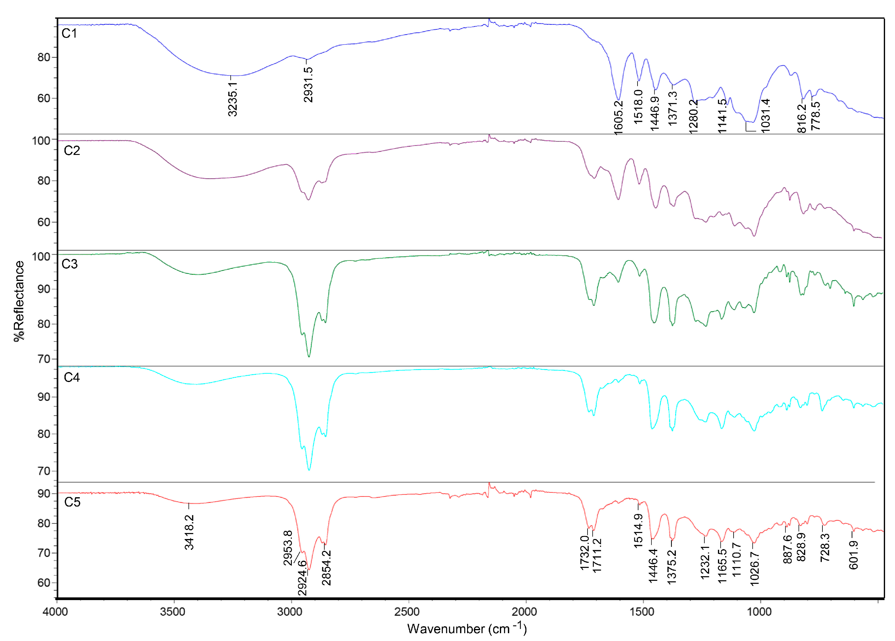
2.2.2. Fourier-Transform Infrared Spectroscopy (FTIR)

Infrared spectra were obtained on a Nicolet iS50 FTIR spectrometer (Thermo Scientific, Waltham, MA, USA) with a diamond single-bounce attenuated total reflectance (ATR) accessory. For each extract, 64 scans were recorded in the 4000–650 cm−1 spectral range in the reflectance mode with a resolution of 4 cm−1. The spectrum of air was used as a background. Spectral data were collected with Omnic v9.2 (Thermo Electron Corp., Waltham, MA, USA) software without post-run processing.

2.3. Antifungal Activity

Antifungal bioassays were carried out based on the Poisoned food method [45] with modifications. For this purpose, malt extract culture medium (MEA: malt extract 1.3%, glucose 1%, and agar 2%) was prepared, and it was sterilized in an autoclave at 130 °C and 1 atm for 20 min. Then, the extract of each solvent was added to the medium dissolved in DMSO (8.3 µL/mL), and 15 mL of MEA was added to the 90 mm Petri dish. The antifungal activity of 0.5 µg/µL of each extract was evaluated against the three fungi: Ch. globosum, T. trogii, and P. sanguineus.
A negative control (C) was prepared by dissolving DMSO without extract in MEA culture medium, and a vitality control of the fungus in MEA without DMSO or extracts (positive control) was also carried out. Petri dishes were inoculated with a 7-day-old plug of a pure fungal isolate of P. sanguineus, T. trogii, and Ch. globosum. Triplicates were made for all the assays in order to reduce the error. Petri dishes were incubated at 26 °C ± 2 °C and a relative humidity of 65%. After six days, the diameter of the colony was measured with a caliper. Antifungal assays were carried out with methanol, acetone, ethyl acetate, and ethyl acetate previously extracted with hexane and dichloromethane extracts.
The percentage of inhibition was calculated as indicated below (Equation (1)) [45]:
i n h i b i t i o n   % = ( C T ) C × 100
where C is the diameter of the colony of the negative control and T is the diameter of the colony of the treatment with each extract.

Statistical Analysis

We employed a linear model to examine the variability in the percentage of inhibition, considering both the type of fungus and the extractives. Additionally, we accounted for the interaction effect between these factors. The statistical analysis was carried out using analysis of variance (ANOVA). Tukey post hoc tests were employed to identify specific groups with different means when statistical significance was observed. A significance level of 0.05 (alpha = 0.05) was used throughout the analysis. All statistical procedures were performed using R [46] and Rstudio software [47].

3. Results and Discussion

3.1. Wood Extraction and Characterization of Extractives

As shown in Table 1, the extraction solvent affects the yields of extractives, which varied from 0.3% to 2.5% for hexane and acetone, respectively. The highest amounts of total extractives were obtained with acetone and ethyl acetate, 250.2 and 184.4 mg/g, respectively. The high variability of the extract concentrations obtained for each solvent is attributed to the diversity of compounds contained in each extractive, which suggests that the non-polar solvent, hexane, may not be the most appropriate option for this wood.
There are no bibliographic records of compounds obtained from the heartwood of C. fissilis. Our results revealed that the percentages obtained from the heartwood extracts were in the range of data previously published for other species of the Cedrela genus [48,49,50]. While the percentages of extractives vary significantly among different types of wood, they usually do not exceed five percent of dry wood mass on average (including heartwood and sapwood), except for some tropical and subtropical woods [51].
The TLC profiles exhibited a complex spot pattern for the extracts, showing similarities between extractives of similar polarity. In this sense, the spot profiles obtained for methanol (C2) and acetone (C3) extracts were very similar, and the same was observed for ethyl acetate (C4) and dichloromethane (C5). This observation was consistent with the results obtained from FTIR-ATR spectra (Figure 1), showing more similarity between C4 and C5 and C2 and C3, respectively, with notable differences compared to the water extract spectrum. Characteristic strong bands in the 3200–3400 cm−1 range may be assigned to the O-H stretching vibrations of hydrogen-bonded hydroxyl groups. Those, together with bands in the 1026–1031 cm−1 range related to the C-OH stretching vibration and other bands in the nearby region characteristic of C-O-C, are probably due to polar compounds, such as low-molecular-weight carbohydrates and polyphenols, which are water extractable constituents of wood [52]. In the methanol extract spectrum, it is possible to observe these bands due to the solubility of said molecules in the solvent. As the polarity of the solvent used decreases, there is a decrease in the intensity of these bands. Also, the bands at 1605, 1446, and 1518 cm−1, possibly representing the aromatic ring (C=C), present differences between the extracts from water and methanol compared to those from other solvents. The intensity of the bands at 2953, 2924, and 2854 cm−1, attributed to methyl and methylene stretching vibrations (C-H), is much greater in extracts C3, C4, and C5. Other characteristics of these extracts are the presence of strong absorption bands at 1711 and 1732 cm−1, consistent with C=O stretching vibrations of esters, aldehydes, ketones, and carboxylic acids (possibly related to the presence of sesquiterpenes and terpenoids, including limonoids, among others compounds previously reported in the genus Cedrela [40]).

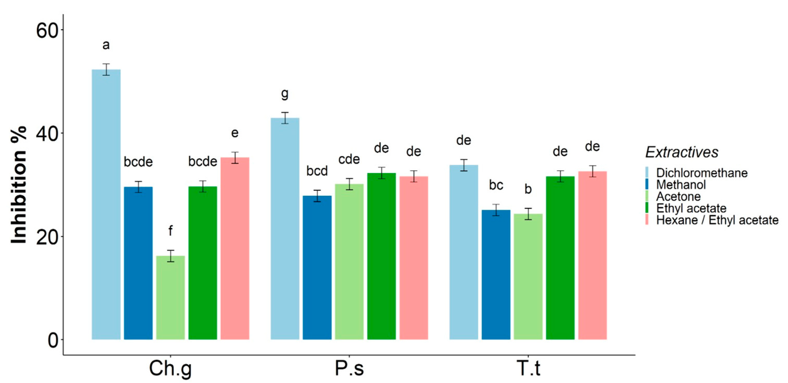
3.2. Antifungal Activity

For all fungi tested after seven days, fungal growth diameters in the media supplemented with the extractives (C2, C3, C4, C5, and C7) were smaller than the respective controls © (Figure 2). Negative controls with DMSO were similar to the viability control without DMSO (positive controls).
In the case of hexane extractives (C6), due to the limited extract obtained and the fact that it was insoluble in DMSO, its antifungal activity was not evaluated. Instead, the antifungal activity of an ethyl acetate extract obtained after extraction with hexane (C7) was evaluated (hexane/ethyl acetate) in order to obtain information about the activity of the extracted compounds with hexane. While prelaminar studies of water extractives (C1) showed that in antifungal tests, the fungal growth was higher than the controls, taking into account that the aim of this work is the search for extractives with antifungal activity, this extract was not further evaluated.
All tested extractives showed inhibition activity to Ch. globosum, P. sanguineus, and T. trogii (Table 2). Considering that the activity of extractives of ethyl acetate (C4) and hexane/ethyl acetate (C7) are very similar, it is possible to propose that the antifungal activity is not directly related to the slightly polar compounds that are extracted with hexane. It is important to highlight the antifungal activity achieved with the dichloromethane extract (C5) against the xylophagous fungi tested in this study. A meaningful inhibition percentage for Ch. globosum was also observed.
Considering that C. fissilis is naturally resistant to fungal attack, our results point towards a possible synergism between the compounds present in the extractives of different polarities. It has been proposed that the level of inhibition of the extractives is related to the extraction solvent, the concentration of the extractives, and the fungal species tested [21,53]. This inhibitory activity could be due to the joint action of compounds rather than the action of a single substance [19,54]. In this sense, Moore et al. [55] studied the effect of the polarity of extractives on the durability of wood and concluded that there are combinations of chemicals in the non-polar and polar components that play an important role.
Studies on the antifungal activity of crude extracts of Cedrela fissilis are very scarce. In the work by Bueno et al., the authors evaluated the antifungal activity of extracts from different parts of the plant Cedrela fissilis against a leafcutter ant and its symbiont fungus. They observed an inhibitory effect of crude dichloromethane extracts of Cedrela fissilis root on the symbiont fungus of the leafcutter ant, with the most effective concentration of the extract being 300 µg/µL [43]. The present results show that at much lower concentrations, such as 0.5 µg/µL of extractive, antifungal activity was observed for the three xylophagous fungi tested: Ch. globosum, T. trogii, and P. sanguineus. This promissory result warrants a future study to determine other inhibitory concentrations.

3.3. Statistical Analysis

The inhibition % was calculated with the measure of the diameter of the fungi, applying Equation (1) and carrying out statistical tests. Initially, to assess variations in the percentage of inhibition, we examined whether both the type of fungus and the type of extract exhibited a relationship with the growth on the dish. An ANOVA test showed there was significant interaction between fungi and extractive, indicating that there was an extractive effect, but its inhibitory activity varied depending on the type of fungus.
After this result, a media comparison was made with the aim of evaluating all combinations between fungi and extractives (Figure 3).
Dichloromethane (C5) extract was the most active for Chaetomium globosum and Pycnoporus sanguineus. Acetone (C3) and methanol (C2) extracts were less effective for Ch. globosum and T. trogii. Even though C5 was less effective for Trametes trogii, it should be noted that the activity with ethyl acetate (C4) and hexane/ethyl acetate (C7) extracts was notable, considering that it is one of the most aggressive white rot fungi of wood. There are reports of antifungal activity of wood extractives against the Trametes genus that are numerous for Trametes versicolor [36,56,57,58], but there are no reports for the antifungal activity of wood extractives for T. trogii, so the results obtained in this work are novel.
It has been described that Cedrela odorata compounds present a biocidal action due to tujaplicin, β-tujaplicinol, and plicatic acid, with antifungal activities against Coniophora puteana, Postia placenta, and Trametes versicolor with metal chelating characteristics [59].
Examples of other wood extractive activities reported that both bark (Acacia mollissima) and heartwood (Schinopsis lorentzii) samples displayed antifungal activity against white rot fungi Trametes versicolor and Pleurotus ostreatus [60], possibly related with their high tannin content [61,62].
Although there are works on the antifungal activity of wood extractives against Pycnoporus sanguineus [36,63,64,65,66], studies against Chaetomium globosum are very scarce. Specifically, for the genus Cedrela, one study reports resistance to the decay by P. sanguineus [67], although this work did not evaluate the extractive activity of this xylophagous fungus. For Ch. globosum, Salem et al. evaluated the antifungal activity of extracts from the heartwood of Pinus rigida, Eucalyptus camaldulensis leaves, and Costus speciosus rhizomes. They observed that extracts from the heartwood of P. rigida showed the greatest activity [68].
Fazio et al. (2010) isolated Ch. globosum from a Jesuit South American polychrome wood sculpture, and the authors observed a high production of cellulases, as well as a degradation pattern showing secondary cell wall cavities typical of soft rot. Moreover, Ch. globosum was found in building materials, which produce highly cytotoxic chaetomins and chaetoglobosins that inhibit cell division and glucose transport [68].
Studies of wood extractive activity against Ch. globosum, such as the results presented in this paper, are relevant considering it is one of the main species responsible for the biodeterioration of Cultural Heritage [69].

4. Conclusions

Although, in recent years, the use of natural biocides as antifungal agents for “opportunistic” fungi has increased, the search for antifungals against xylophagous-type fungi that attack the Cultural Heritage remains an understudied field.
The importance of this study lies in the fact that Cedrela fissilis, also called “Ygari” or “Cedro misionero”, is a wood that was widely used as one of the Guaraní Jesuit materials in the 17th and 18th centuries; hence, it is important to know its capacity to resist fungi of wood rot. The knowledge of extractives of wood and their antifungal activity generates relevant information to better understand and harness the properties of each wood. This work contributes to the knowledge of Cedrela fissilis, which is a scarcely studied species.
The present work represents an interesting precedent on the evaluation of wood extractives obtained through a simple protocol, revealing the potential of hardwood extractives as sources of biocides.
Future studies are necessary in order to investigate the obtention of natural compounds from wood waste as an environmentally friendly source of biocides against xylophagous fungi that cause wood rot, which represents one of the main problems for Cultural Heritage.

Author Contributions

Conceptualization, V.P.C. and A.T.F.; methodology, C.S.V., V.P.C. and A.T.F.; formal analysis, C.S.V., T.G.G., V.P.C. and A.T.F.; investigation, C.S.V., V.P.C. and A.T.F.; resources, V.P.C. and A.T.F.; data curation, C.S.V., V.P.C. and A.T.F.; writing—original draft preparation, C.S.V., V.P.C. and A.T.F.; writing—review and editing, V.P.C. and A.T.F.; visualization, C.S.V., T.G.G., V.P.C. and A.T.F.; supervision, V.P.C. and A.T.F.; project administration, V.P.C. and A.T.F.; funding acquisition, V.P.C. and A.T.F. All authors have read and agreed to the published version of the manuscript.

Funding

This research was funded by the Agencia Nacional de Promoción Científica y Tecnológica (ANPCyT) (PICT 2019-1000), the Consejo Nacional de Investigaciones Científicas y Técnicas (CONICET) (PIP 11220170100283CO, PIP 11220200100811CO, and PIBBA 2022–2023: 2872021010 0451CO), and the University of Buenos Aires (20020220100168BA). V.P.C. and A.T.F. are Research Members of CONICET.

Institutional Review Board Statement

Not applicable.

Informed Consent Statement

Not applicable.

Data Availability Statement

Data are contained within the article.

Acknowledgments

C.S.V. thanks the Universidad de Buenos Aires for a Doctoral Fellowship. The authors thank BAFC: the Mycological Culture Collection of the Department of Biological Sciences, Faculty of Exact and Natural Sciences, University of Buenos Aires. We thank Irene Lantos for language editing.

Conflicts of Interest

The authors declare no conflicts of interest.

References

  1. Blanchette, R.A. A review of microbial deterioration found in archaeological wood from different environments. Int. Biodeterior. Biodegrad. 2000, 90, 93–98. [Google Scholar] [CrossRef]
  2. Ciferri, O. The role of microorganisms in the degradation of cultural heritage. Stud. Conserv. 2002, 47, 35–45. [Google Scholar] [CrossRef]
  3. Strzelczyk, A.B. Observations on aesthetic and structural changes induced in Polish historic objects by microorganisms. Int. Biodeterior. Biodegrad. 2004, 53, 151–156. [Google Scholar] [CrossRef]
  4. Pournou, A. Biodeterioration of Wooden Cultural Heritage: Organisms and Decay Mechanisms in Aquatic and Terrestrial Ecosystems; Springer: Cham, Swizerland, 2020; 538p. [Google Scholar] [CrossRef]
  5. Fernández Fueyo, E.; Ruiz Dueñas, F.J.; López Lucendo, M.F.; Pérez Boada, M.; Rencoret, J.; Gutierrez, A.; Pisabarro, A.G.; Ramirez, L.; Martinez, A.T. A secretomic view of woody and nonwoody lignocellulose degradation by Pleurotus ostreatus. Biotechnol. Biofuels 2016, 9, 49. [Google Scholar] [CrossRef] [PubMed]
  6. Naidu, Y.; Siddiqui, Y.; Rafii, M.Y.; Saud, H.M.; Idris, A.S. Investigating the effect of white-rot hymenomycetes biodegradation on basal stem rot infected oil palm wood blocks: Biochemical and anatomical characterization. Ind. Crops Prod. 2017, 108, 872–882. [Google Scholar] [CrossRef]
  7. Levin, L.N.; Castro, M.A. Anatomical study of the decay caused by the white-rot fungus Trametes trogii (Aphyllophorales) in wood of Salix and Populus. IAWA J. 1998, 19, 169–180. [Google Scholar] [CrossRef]
  8. Levin, L.; Viale, A.; Forchiassin, A. Degradation of organic pollutants by the white rot basidiomycete Trametes trogii. Int. Biodeterior. Biodegrad. 2003, 52, 1–5. [Google Scholar] [CrossRef]
  9. Luna, M.; Murace, M.; Keil, G.; Otaño, M. Patterns of decay caused by Pycnoporus sanguineus and Ganoderma lucidum (Aphyllophorales) in poplar wood. IAWA J. 2004, 25, 425–433. [Google Scholar] [CrossRef]
  10. Nilsson, T. Studies on wood degradation and cellulolytic activity of microfungi. Stud. For. Suec. 1973, 104, 1–39. [Google Scholar]
  11. Eaton, R.A.; Hale, M.D.C. Wood: Decay, Pests and Protection; Chapman & Hall: London, UK, 1993. [Google Scholar]
  12. Wozniak, M. Antifungal agents in wood protection. A review. Molecules 2022, 27, 6392. [Google Scholar] [CrossRef]
  13. Onuorah, E.O. The wood preservative potentials of heartwood extracts of Milicia excela and Erthrophleum suaveolens. Bioresour. Technol. 2000, 75, 171–173. [Google Scholar] [CrossRef]
  14. Mbakidi-Ngouaby, H.; Pinault, E.; Gloaguen, V.; Costa, G.; Sol, V.; Millot, M.; Mambu, L. Profiling and seasonal variation of chemical constituents from Pseudotsuga menziesii wood. Ind. Crops Prod. 2018, 117, 34–49. [Google Scholar] [CrossRef]
  15. Singh, T.; Singh, A.P. A review on natural products as wood protectant. Wood Sci. Technol. 2012, 46, 851–870. [Google Scholar] [CrossRef]
  16. Lagrouh, F.; Dakka, N.; Bakri, Y. The antifungal activity of Moroccan plants and the mechanism of action of secondary metabolites from plants. J. Mycol. Med. 2017, 27, 303–311. [Google Scholar] [CrossRef] [PubMed]
  17. Brocco, V.F.; Paes, J.B.; Costa, L.G.; da Brazolin, S.; Arantes, M.D.C. Potential of teak heartwood extracts as a natural wood preservative. J. Clean. Prod. 2017, 142, 2093–2099. [Google Scholar] [CrossRef]
  18. Kadir, R.; Hale, M. Biocidal potential of the extractives of four Malaysian timbers against subterranean termites and wood decay fungi. Eur. J. Wood Prod. 2019, 77, 147–155. [Google Scholar] [CrossRef]
  19. Broda, M. Natural Compounds for Wood Protection against Fungi. A review. Molecules 2020, 25, 3538. [Google Scholar] [CrossRef]
  20. Eller, F.J.; Kirker, G.T.; Mankowski, M.E.; Hay, W.T.; Palmquist, D.E. Effect of burgundy solid extracted from Eastern Red Cedar heartwood on subterranean termites and Wood-decay fungi. Ind. Crops Prod. 2020, 144, 112023. [Google Scholar] [CrossRef]
  21. Wu, J.F.; Zhuang, X.F.; Yuan, H.M.; Li, Q.; Wang, W.; Li, J.Q.; Lin, J.G. Antifungal ability, and decay resistance of Fokienia hodginsii heartwood extract and its inhibitory effect on Gloeophyllum trabeum. BioResources 2019, 15, 2784–2799. [Google Scholar] [CrossRef]
  22. Santos, M.B.; Sillero, L.; Gatto, D.A.; Labidi, J. Bioactive molecules in wood extractives: Methods of extraction and separation, a review. Ind. Crops Prod. 2022, 186, 115231. [Google Scholar] [CrossRef]
  23. Rowell, R.M.; Pettersen, R.; Tshabalala, M.A. Cell Wall Chemistry. In Handbook of Wood Chemistry and Wood Composites, 2nd ed.; Roger, M., Ed.; Rowell CRC Press: Boca Raton, FL, USA, 2012; pp. 34–70. [Google Scholar]
  24. Doussot, F.; De Jéso, B.; Quideau, S.; Pardon, P. Extractives content in cooperage oak wood during natural seasoning and toasting; influence of tree species, geographic location, and single-tree effects. J. Agric. Food Chem. 2002, 50, 5955–5961. [Google Scholar] [CrossRef]
  25. Kebbi Benkeder, Z.; Colin, F.; Dumarçay, S.; Gérardin, P. Quantification and characterization of knotwood extractives of 12 European softwood and hardwood species. Ann. For. Sci. 2014, 72, 277–284. [Google Scholar] [CrossRef]
  26. Prida, A.; Puech, J.L. Influence of geographical origin and botanical species on the content of extractives in American, French, and East European oak woods. J. Agric. Food Chem. 2006, 21, 8115–8126. [Google Scholar] [CrossRef]
  27. Taylor, A.M.; Gartner, B.L.; Morrell, J.J. Heartwood formation and natural durability. A review. Wood Fiber Sci. 2002, 34, 587–611. [Google Scholar]
  28. Kilpeläinen, A.; Peltola, H.; Ryyppö, A. Scots pine responses to elevated temperature and carbon dioxide concentration: Growth and wood properties. Tree Physiol. 2005, 25, 75–83. [Google Scholar] [CrossRef]
  29. Achim, A.; Blanchet, P.; Schmitt, U.; Bouffard, J.F. Characterization of extractives in tree biomass and by-products of Plywood and Saw Mills from Finnish birch in different climatic regions for value-added chemical products. In Proceedings of the ISCHP 15: International Scientific Conference on Hardwood Processing Proceedings, Québec City, QC, Canada, 15–17 September 2015. [Google Scholar]
  30. Routa, J.; Brännström, H.; Anttila, P.; Mäkinen, M.; Jänis, J.; Asikainen, A. Wood extractives of Finnish pine, spruce and birch–availability and optimal sources of compounds: A literature review. Nat. Resour. Bioecon. Stud. 2017, 73, 55. [Google Scholar]
  31. Challinor, J.M. Characterization of wood extractives by pyrolysis-gas chromatography/mass spectrometry of quaternary ammonium hydroxide extracts. J. Anal. Appl. Pyrolysis 1996, 37, 1–13. [Google Scholar] [CrossRef]
  32. Royer, M.; Herbette, G.; Eparvier, V.; Beauchêne, J.; Thibaut, B.; Stien, D. Secondary metabolites of Bagassa guianensis Aubl. wood: A study of the chemotaxonomy of the Moraceae family. Phytochem 2010, 71, 1708–1713. [Google Scholar] [CrossRef]
  33. Yin, X.; Huang, A.; Zhang, S.; Liu, R.; Ma, F. Identification of three Dalbergia species based on differences in extractive components. Molecules 2018, 23, 2163. [Google Scholar] [CrossRef]
  34. Felhofer, M.; Prats-Mateu, B.; Bock, P.; Gierlinger, N. Antifungal stilbene impregnation: Transport and distribution on the micron-level. Tree Physiol. 2018, 38, 1526–1537. [Google Scholar] [CrossRef] [PubMed]
  35. Valette, N.; Perrot, T.; Sormani, R.; Gelhaye, E.; Morel Rouhier, M. Antifungal activities of wood extractives. Fungal Biol. Rev. 2017, 31, 113–123. [Google Scholar] [CrossRef]
  36. Anouhe, J.B.S.; Niamké, F.B.; Faustin, M. The role of extractives in the natural durability of the heartwood of Dicorynia guianensis Amsh: New insights in antioxydant and antifungal properties. Ann. For. Sci. 2018, 75, 15. [Google Scholar] [CrossRef]
  37. Ateş, S.; Gür, M. Chemical contents and antifungal activity of some durable wood extractives vs. Pleurotus ostreatus. BioResources 2015, 10, 2433–2443. [Google Scholar] [CrossRef]
  38. Wang, J.; Li, J.; Li, S.; Freitag, C.; Morrell, J.J. Antifungal activities of Cunninghamia lanceolata heartwood extractives. BioResources 2011, 6, 606–614. [Google Scholar] [CrossRef]
  39. de Andrade, J.H.C.; Rodrigues, J.; Benites, A.; Benites, C.; Acosta, A.; Benites, M.; Benites, C.; Gomes, I.; da Silva, J.V.; Antunes, E.; et al. Notes on current Mbyá-Guarani medicinal plant exchanges in southern Brazil. J. Ethnobiol. Ethnomed. 2021, 17, 38. [Google Scholar] [CrossRef]
  40. Nogueira, T.S.R.; Passos, M.S.; Nascimento, L.P.S.; Arantes, M.B.S.; Monteiro, N.O.; Bueno, S.I.D.S.; de Carvalho Junior, A.; Azevedo, O.A.; Terra, W.D.S.; Vieira, M.G.C.; et al. Chemical compounds and biologic activities: A Review of Cedrela genus. Molecules 2020, 25, 5401. [Google Scholar] [CrossRef]
  41. Ambrozin, A.R.; Leite, A.C.; Bueno, F.C.; Vieira, P.C.; Fernandes, J.B.; Bueno, O.C.; Bacci, M., Jr. Limonoids from andiroba oil and Cedrela fissilis and their insecticidal activity. J. Braz. Chem. Soc. 2006, 17, 542–547. [Google Scholar] [CrossRef]
  42. Leite, A.C.; Ambrozin, A.R.P.; Fernandes, J.B.; Vieira, P.C.; Da Silva, M.F.D.G.F.; De Albuquerque, S. Trypanocidal activity of limonoids and triterpenes from Cedrela fissilis. Planta Med. 2008, 74, 1795–1799. [Google Scholar] [CrossRef]
  43. Bueno, F.C.; Godoy, M.P.; Leite, A.C.; Bueno, O.C.; Pagnocca, F.C.; Fernandes, J.B.; Hebling, M.J.A.; Bacci, M.; Vieira, P.C.; Silva, M.F.G.F. Toxicity of Cedrela fissilis to Atta sexdens rubropilosa (Hymenoptera: Formicidae) and its symbiotic fungus. Sociobiology 2005, 45, 389–399. [Google Scholar]
  44. Fazio, A.T.; Papinutti, L.; Gómez, A.; Parera, S.D.; Rodríguez Romero, A.; Siracusano, G.; Maier, M.S. Fungal deterioration of a Jesuit South American polychrome wood sculpture. Int. Biodeterior. Biodegrad. 2010, 64, 694–701. [Google Scholar] [CrossRef]
  45. Balouiri, M.; Sadiki, M.; Koraichi Ibnsouda, S. Methods for in vitro evaluating antimicrobial activity: A review. J. Pharm. Anal. 2016, 6, 71–79. [Google Scholar] [CrossRef] [PubMed]
  46. Posit Team. RStudio: Integrated Development Environment for R; Posit Software; PBC: Boston, MA, USA, 2023; Available online: http://www.posit.co/ (accessed on 25 December 2023).
  47. R Core Team. R: A Language and Environment for Statistical Computing; R Foundation for Statistical Computing: Vienna, Austria, 2023; Available online: https://www.R-project.org/ (accessed on 25 December 2023).
  48. Campos, A.M.; Oliveira, F.S.; Machado, M.I.L.; Matos, F.J.A.; Braz-Filho, R. Triterpenes from Cedrela odorata. Phytochem 1991, 30, 1225–1229. [Google Scholar] [CrossRef]
  49. Segura, R.; Calderón, J.; Toscano, R.; Gutiérrez, A.; Mata, R. Cedrelanolide I, A new limonoid from Cedrela salvadorenis. Tetrahedron Lett. 1994, 35, 3427–3440. [Google Scholar] [CrossRef]
  50. Céspedes, C.L.; Calderón, J.S.; Gomez-Garibay, F.; Segura, R.; King-Diaz, B.; Lotina-Hennsen, B. Phytogrowth properties of limonoids isolated from Cedrela ciliolata. J. Chem. Ecol. 1999, 25, 2665–2676. [Google Scholar] [CrossRef]
  51. Lehr, M.; Miltner, M.; Friedl, A. Removal of wood extractives as pulp (pre-)treatment: A technological review. SN Appl. Sci. 2021, 3, 886. [Google Scholar] [CrossRef]
  52. Björklund, J.; Marianne; Nilvebrant, N.O. Wood Chemistry and Wood Biotechnology; De Gruyter: Berlin, Germany, 2009; Volume 1, pp. 147–172. [Google Scholar]
  53. Lv, H.; Wang, Y.; Qu, M.; Zhang, Y.; Song, Z.; Su, X.; Xu, B. Anti-fungal Activity of Dalbergia retusa Extract on Gloeophyllum trabeum. Front. Plant Sci. 2022, 13, 906041. [Google Scholar] [CrossRef] [PubMed]
  54. Ribeiro, S.B.; Pagnocca, F.C.; Victor, S.R.; Bueno, O.C.; Hebling, M.G.; Bacci, M., Jr.; Silva, O.A.; Fernandes, J.B.; Vieira, P.C.; Silva, M.F.G.F. Activity of sesame leaf extracts Against the symbiotic fungus of Atta sexdens L. An. Soc. Entomológica Bras. 1998, 27, 421–426. [Google Scholar] [CrossRef]
  55. Moore, R.K.; Smaglick, J.; Arellano-ruiz, E.; Leitch, M.; Mann, D. The effect of polarity of extractives on the durability of wood. In Proceedings of the 18th ISWFPC (International Symposium on Wood, Fiber, and Pulping Chemistry), Vienna, Austria, 9–11 September 2015. [Google Scholar]
  56. Vek, V.; Oven, P.; Humar, M. Phenolic extractives of wound-associated wood of beech and their fungicidal effect. Int. Biodeterior. Biodegrad. 2013, 77, 91–97. [Google Scholar] [CrossRef]
  57. Tchinda, J.B.S.; Ndikontar, M.K.; Founda Belinga, A.D.; Mounguengui, S.; Njankouo, J.M.; Durmaçay, S.; Geradin, P. Inhibition of fungi with wood extractives and natural durability of five Cameroonian wood species. Ind. Crops Prod. 2018, 123, 183–191. [Google Scholar] [CrossRef]
  58. Candelier, K.; Thévenon, M.F.; Collet, R.; Gérardin, P.; Dumarçay, S. Antifungal and antitermite activity of extractives compunds from thermally modified ash woods. Maderas. Cienc. Tecnol. 2020, 22, 223–240. [Google Scholar] [CrossRef]
  59. Stirling, R.; Daniels, C.R.; Clark, J.E.; Morris, P.I. Methods for Determining the Role of Extractives in the Natural Durability of Western Red Cedar; IRG: Stockholm, Sweden, 2007. [Google Scholar]
  60. Tascioglu, C.; Yalcin, M.; Sen, S.; Akcay, C. Antifungal properties of some plant extracts used as wood preservatives. Int. Biodeterior. Biodegrad. 2013, 85, 23–28. [Google Scholar] [CrossRef]
  61. Roux, D.G.; Ferreira, D.; Botha, J.J. Structural considerations in predicting the utilization of tannins. J. Agric. Food Chem. 1980, 28, 216–222. [Google Scholar] [CrossRef]
  62. Roux, D.G. Reflections on the chemistry and affinities of the major commercial condensed tannins in the context of their industrial use. In Plant Polyphenols; Basic Life Sciences; Springer: Boston, MA, USA, 1992. [Google Scholar] [CrossRef]
  63. Kawamura, F.; Mahamud, A.; Sulaiman, O.; Hashim, R. Antifungal activities of extracts from heartwood, sapwood and bark of 11 malaysian timbers against Gloeophyllum trabeum and Pycnoporus sanguineus. J. Trop. For. Sci. 2010, 22, 170–174. [Google Scholar]
  64. Anis, Z.; Sulaiman, O.; Hashim, R.; Mehdi, S.H.; Ghali, R.M. Radical scavenging activity, total phenol content and antifungal activity of Cinnamomum iners wood. Iran. J. Energy Environ. 2012, 3, 74–78. [Google Scholar] [CrossRef]
  65. Bento, T.S.; Torres, L.M.B.; Fialho, M.B.; Bononi, V.L.R. Growth inhibition and antioxidative response of wood decay fungi exposed to plant extracts of Casearia species. Lett. Appl. Microbiol. 2013, 58, 79–86. [Google Scholar] [CrossRef] [PubMed]
  66. Harmen, M.S.; Ramle, S.F.M. Antifungal activities of methanol extract from Syzygium chlorantha and Hopea spp. against Pycnoporus sanguineus. J. Trop. Resour. Sustain. 2016, 4, 88–94. [Google Scholar] [CrossRef]
  67. Souza Reis, A.R.; Gomes Loch, A.; da Costa Oliveira Moreir, S.M.; da Costa Gondim, M.; Rogério da Silva, M.; Soares Bilhalva dos Santos, P. Natural resistance of wood from three forest species exposed to Pycnoporus sanguineus xylophagous fungus. Adv. For. Sci. 2019, 6, 839–842. [Google Scholar] [CrossRef]
  68. Salem, M.Z.; Zidan, Y.E.; El Hadidi, N.M.; Mansour, M.M.; Elgat, W.A.A. Evaluation of usage three natural extracts applied to three commercial wood species against five common molds. Int. Biodeter. Biodegr. 2016, 110, 206–226. [Google Scholar] [CrossRef]
  69. Cirone, M.; Figoli, A.; Galiano, F.; La Russa, M.F.; Macchia, A.; Mancuso, R.; Ricca, M.; Rovella, N.; Taverniti, M.; Ruffolo, S.A. Innovative methodologies for the conservation of cultural heritage against biodeterioration: A review. Coatings 2023, 13, 1986. [Google Scholar] [CrossRef]
Figure 1. FTIR-ATR spectra of extractives: C1 (water), C2 (methanol), C3 (acetone), C4 (ethyl acetate), C5 (dichloromethane). FTIR conditions: number of scans (64) and resolution (4 cm−1).
Coatings 14 00237 g001
Figure 2. Petri dishes with fungus over 7 days of growth. Assays with extractives of methanol (C2), acetone (C3), ethyl acetate (C4), dichloromethane (C5), ethyl acetate extract obtained after extraction with hexane (C7), and negative controls with DMSO. Positive controls: Chaetomium globosum (Ch.g), Pycnoporus sanguineus (P.s), and Trametes trogii (T.t) without DMSO.
Coatings 14 00237 g002
Figure 3. Estimated means by fungi and extractives. Values are shown as estimated mean (±standard error). Different letters indicate there are significant differences between groups (confidence level used: 0.95, Tukey’s post hoc comparison test).
Coatings 14 00237 g003
Table 1. Extractives C1–C7 (C1: water, C2: methanol, C3: acetone, C4 ethyl acetate, C5: dichloromethane, C6: hexane, C7: ethyl acetate extract obtained after extraction with hexane). Extraction method: initial mass (10 g) of C. fissilis, extraction solvent (150 mL), and maceration at room temperature 48 h.
Extract CodeExtract Recovered (mg)% Extract Recovered
C153.50.5
C271.90.7
C3250.22.5
C4184.41.8
C5100.21
C628.60.3
C774.80.7
Table 2. Inhibition % of extractives of Cedrela fissilis after seven days of incubation. Values are mean ± standard deviation for bioassays conducted in triplicate.
Inhibition (%)
Extract CodeCh. globosumP. sanguineusT. trogii
C229.57 ± 1.5627.84 ± 2.2325.12 ± 2.93
C316.20 ± 3.9530.11 ± 1.6624.34 ± 2.00
C429.66 ± 2.2932.28 ± 1.0831.62 ± 1.93
C552.31 ± 1.7342.92 ± 1.5633.77 ± 2.76
C735.22 ± 2.4831.62 ± 0.8032.61 ± 1.60
Disclaimer/Publisher’s Note: The statements, opinions and data contained in all publications are solely those of the individual author(s) and contributor(s) and not of MDPI and/or the editor(s). MDPI and/or the editor(s) disclaim responsibility for any injury to people or property resulting from any ideas, methods, instructions or products referred to in the content.

