Article

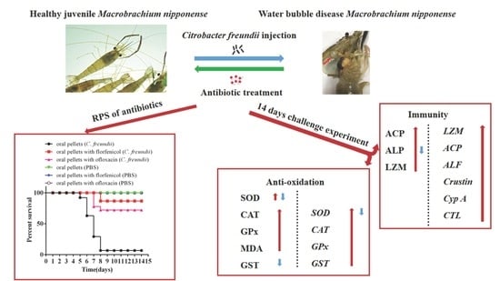
A Novel Disease (Water Bubble Disease) of the Giant Freshwater Prawn Macrobrachium rosenbergii Caused by Citrobacter freundii: Antibiotic Treatment and Effects on the Antioxidant Enzyme Activity and Immune Responses

1
State Key Laboratory for Biocontrol, School of Marine Sciences, Sun Yat-sen University, No.132 Waihuan Dong Road, Higher Education Mega Center, Guangzhou 510006, China
2
Southtern Marine Science and Engineering Guangdong Laboratory (Zhuhai), School of Life Science, Sun Yat-sen University, No.132 Waihuan Dong Road, Higher Education Mega Center, Guangzhou 510006, China
*
Author to whom correspondence should be addressed.
Antioxidants 2022, 11(8), 1491; https://doi.org/10.3390/antiox11081491
Submission received: 14 June 2022 / Revised: 23 July 2022 / Accepted: 26 July 2022 / Published: 29 July 2022
(This article belongs to the Special Issue Oxidative Stress in Aquatic Organisms)

Abstract

:
The giant freshwater prawn, Macrobrachium rosenbergii, is an important and economical aquaculture species widely farmed in tropical and subtropical areas of the world. A new disease, “water bubble disease (WBD)”, has emerged and resulted in a large loss of M. rosenbergii cultured in China. A water bubble with a diameter of about 7 mm under the carapace represents the main clinical sign of diseased prawns. In the present study, Citrobacter freundii was isolated and identified from the water bubble. The optimum temperature, pH, and salinity of the C. freundii were 32 °C, 6, and 1%, respectively. A challenging experiment showed that C. freundii caused the same typical signs of WBD in prawns. Median lethal dose of the C. freundii to prawn was 104.94 CFU/g. According to the antibiogram tests of C. freundii, florfenicol and ofloxacin were selected to evaluate their therapeutic effects against C. freundii in prawn. After the challenge with C. freundii, 86.67% and 72.22% survival of protective effects against C. freundii were evaluated in the oral florfenicol pellets and oral ofloxacin pellets feding prawns, respectively, whereas the mortality of prawns without fed antibiotics was 93%. After antibiotic treatment and C. freundii infection, the activities of superoxide dismutase (SOD), catalase (CAT), glutathione peroxidase (GPx), glutathione S-transferase (GST), malondialdehyde (MDA), acid phosphatase (ACP), alkaline phosphatase (ALP), and lysozyme (LZM) in the hemolymph and hepatopancreas of the prawns and the immune-related gene expression levels of Cu/Zn-SOD, CAT, GPx, GST, LZM, ACP, anti-lipopolysaccharide factor, crustin, cyclophilin A, and C-type lectin in hepatopancreas were all significantly changed, indicating that innate immune responses were induced by C. freundii. These results can be beneficial for the prevention and control of C. freundii in prawns.

Graphical Abstract

1. Introduction

The giant freshwater prawn, Macrobrachium rosenbergii, is a member of arthropods, belonging to genus Macrobrachium, which is the largest freshwater and the most favored prawn for aquaculture in tropical and subtropical areas of the world [1,2,3], with annual yields of 290,708 metric tons [4]. M. rosenbergii has already been developed into an important economically aquaculture species in China [5,6,7,8]. The outbreaks of bacterial diseases in M. rosenbergii aquaculture have been reported, such as muscle necrosis disease caused by Enterococcus or Pseudomonas aeruginosa [9,10,11], Aeromonas species (A. veronii and A. caviae) and Vibrio species (V. alginolyticus and V. parahaemolyticus) [12,13] caused high-mortality diseases. A new epidemic disease, referred to as “water bubble disease” (WBD), recently occurred in cultured M. rosenbergii in China, and it caused a mortality of over 30% in diseased prawns.
As M. rosenbergii is an invertebrate, it lacks the typical acquired immunity of vertebrates and entirely relies on its innate immunity system to resist infection by various pathogens [14]. Enhanced knowledge about the immune system of M. rosenbergii is crucial for disease management [15]. The innate immune system provides defense against invading bacteria through AMPs, enzymes, and cellular components [16]. Antiproteases, myeloperoxidase, and lysozyme (LZM) have been used to learn about and determine the innate immune status of an organism [17]. During pathogen invasion, host cells produce abundant reactive oxygen species (ROS) to kill the invading pathogens. However, a considerably high level of ROS in the body will cause the destruction and damage of DNA and other biological macromolecules [18]. Several major antioxidant enzymes include glutathione peroxidase (GPx), glutathione S-transferase (GST), catalase (CAT), malondialdehyde (MDA), and superoxide dismutase (SOD), which play important roles in anti-oxidization damage and related coping mechanisms [19,20]. Acid phosphatase (ACP), and alkaline phosphatase (ALP) are symbols of macrophage activation, important components of the lysosome system, and play important roles in the innate immune system by engulfing antigens and LZM intracellularly [21]. C-Type lectin (CTL), cyclophilin A (CypA), and AMPs, such as anti-lipopolysaccharide factor (ALF) and crustins, play important roles in the innate immunity for the recognition of effector molecules and clearance of bacteria, fungi, and viruses [22,23,24,25,26,27,28,29]. Antibiotics are commonly used in aquaculture to control the diseases caused by bacteria [30]. Antibiotics at therapeutic levels are mainly administered for short periods of time via the oral route (the most common route for the delivery occurs by mixing the antibiotic with commercial feed) to groups of prawn. Antibiotics legally used in aquaculture must be authorized by Food and Drug Administration (FDA), and abide by the rules for antibiotic use, comprising permissible routes of delivery, dose forms, withdrawal times, tolerances, and dose rates and limitations of specific species. In Norway, antibiotics were sold in pharmacies or in feed plants approved by the Norwegian Medicines Agency, and the amount of antibiotics used and retain records in prescriptions of veterinarian were mandatory to report [31].
In the present study, we aimed to isolate and identify the pathogen associated with WBD in M. rosenbergii using challenge experiments, molecular methods and antibiotic treatment. The activities of antioxidant enzymes (i.e., SOD, CAT, GPx, GST, and MDA), ACP, ALP, and LZM and the immune-related gene expression levels of Cu/Zn-SOD, CAT, GPx, GST, LZM, ACP, ALF, crustin, CypA, and CTL in M. rosenbergii after C. freundii challenge were investigated. The results of the present study will expound the pathogenesis of bacteria in M. rosenbergii and facilitate the further prevention and control of C. freundii in M. rosenbergii.

2. Materials and Methods

2.1. Strain Isolation and Identification

In November 2019, 30 M. rosenbergii (mean body weight 13.5 ± 2.3 g, body length 13.1 ± 1.2 cm) with WBD were collected in Zhaoqing, Guangdong, China. The water bubble diseased M. rosenbergii were dissected aseptically, and liquid sample of the water bubble was streaked onto a Luria-Bertani (LB) agarose plate (tryptone 10 g/L, yeast extract 5 g/L, NaCl 10 g/L, agarose powder 15 g/L, pH 7.4) (Sangon Biotech, Shanghai, China). The inoculated plates were incubated for 48 h at 28 °C, and single colonies were obtained and labeled GDZQ201912. After incubation in liquid LB culture (tryptone 10 g/L, yeast extract 5 g/L, NaCl 10 g/L, pH 7.4) for 24 h, the bacteria were collected and stored at −80 °C in 50% glycerol (v/v).
Biochemical characterization of the isolated strain was identified by Gram staining, shape, and motility, followed by biochemical tests and carbohydrate utilization test. The biochemical tests were performed with GEN III MicroPlate on BIOLOGY (Biology, Vacaville, CA, USA). Transmission electron microscopy (TEM) (JEOL JEM-1400, JEOL, Tokyo, Japan) was used to observe the isolated strain at 120 kV.

2.2. Gene Sequencing and Phylogenetic Analysis

Genomic DNA of the bacterial isolate was extracted by using FastPure Cell/Tissue DNA Isolation Mini Kit (Vazyme, Nanjing, China). The 16S rRNA gene of the bacterium was amplified for polymerase chain reaction (PCR) analysis in accordance with the method of Lee et al. [32]. The following amplification primers were used: F (5′-AGAGTTTGATCCTGGCTCAG-3′) and R (5′-CGGTTACCTTGTTACGACTT-3′) (NR_176804.1). The PCR program was carried out as follows: denaturation at 95 °C for 10 min, 30 cycles of denaturation at 95 °C for 30 s, annealing at 53 °C for 30 s, extension at 72 °C for 80 s, and a final extension at 72 °C for 10 min. PCR products were run on a TBE-agarose gel, and the DNA was purified using a TaKaRa MiniBEST Universal Genomic DNA Extraction Kit (Takara, Kusatsu, Japan) and sub-cloned into the pMD19-T Easy Vector (Takara, Japan) for sequencing.
The 1416 bp PCR product was sequenced and blast analyzed. The 16S rRNA gene sequence of GDZQ201912 was aligned with the 16S rRNA gene sequences of other Citrobacter (KC210829.1, FN997639.1, MG011554.1, AP022399.1, AP022486.1, and AP022513.1), Edwardsiella (FJ405305.1, EF467289.1, GQ180182.1, FJ405309.1, GQ180181.1, and KC309472.1), Vibrio (MT071600.1, MT269596.1, MT307282.1, and MT505697.1), and Aeromonas species (EU770272.1, KF358430.1, MK182872.1, and MK182893.1) retrieved from the National Center for Biotechnology Information database. The phylogenetic tree based on 16S rRNA gene sequences of the bacteria cells was constructed via the neighbor-joining (NJ) method with the MEGA program (version 7) using a maximum composite likelihood model. The robustness of the NJ tree was assessed using the approximate likelihood-ratio test and bootstrapping with 1000 replicates. Bootstrap values were shown at each node.

2.3. Effects of Temperature, pH, and Salinity on the Growth of Citrobacter freundii

The isolated strain was inoculated into the LB liquid medium and homogenized. Different temperatures (24 °C, 28 °C, 32 °C, and 42 °C) were used to test their effects on the growth of C. freundii at pH 7.0 and salinity 1%. Different pH (3.0, 4.0, 5.0, 6.0, 7.0, 8.0, 9.0, and 10.0) were used to test their effects on the growth of C. freundii at 28 °C temperature and 1% salinity. Different salinities (0%, 1%, 2.0%, 3%, 4%, 5%, 6%, and 7%) were used to test their effects on the growth of C. freundii at 28 °C temperature and pH 7.0. A 210 µL LB liquid medium containing the isolated strain (102 CFU/mL) of different temperatures, pH, and salinities was added to each well of 96-well plates. Then, the bacteria were sampled regularly in triplicate to determine the optical density at a wavelength of 600 nm (OD600) per 30 min and cultured for 24 h.

2.4. Antibiotic Susceptibility Test

The antibiotic susceptibility test was performed using 106 CFU/mL isolated strain in the disk diffusion method as described by Lalitha [33]. The isolated strain was tested following the antimicrobial agents (disk content indicated in parentheses): penicillin (10 µg), doxycycline (30 µg), florfenicol (30 µg), norfloxacin (10 µg), ofloxacin (5 µg), amoxicillin (20 µg), ampicillin (10 µg), cefepime (30 µg), ceftriaxone (30 µg), neomycin (30 µg), gentamicin (10 µg), streptomycin (10 µg), clindamycin (2 µg), lincomycin (2 µg), tetracycline (30 µg), novobiocin (30 µg), polymyxin B (300 µg), and rifampin (5 µg) (HANGWEI, Beijing, China). The sizes of the inhibition zones were measured and recorded triplicately. The experimental results were judged in accordance with the clinical and laboratory standards institute standards [34].

2.5. Experimental Challenge

Prawns were obtained from an aquaculture farm in Guandong and acclimated in a recirculating-water aquarium system filled with aerated freshwater for a week. The prawns were fed twice per day with commercial pellet feed (Table 1). Water was maintained at 28 ± 1 °C using a water heater. Healthy prawns (11.00 ± 1.38 g) were used in the following challenge tests. The challenge tests included two groups: a control group, in which 30 prawns were injected intramuscularly with 100 µL phosphate-buffered saline (PBS) at the junction between the 3rd and 4th abdominal segments, and an infected group, in which 30 prawns were injected with 100 µL bacteria inocula (106.76 CFU/g) at the same junction. The 30 prawns of the normal or infected group were stocked in three 36 L aquaria (10 animals per aquarium). The survival rates and clinical signs of the infected and control groups were recorded daily.

2.6. Median Lethal Dose Determination

In total, 240 healthy prawns (8.00 ± 0.79 g) were used and stocked in 36 L aquaria (10 animals per aquarium). The C. freundii (108.01 cells/g) were used for the challenge experiments. For each trial, 10 animals were injected intramuscularly with the C. freundii (100 µL) at 10-fold serial dilutions (100–10−6). Animals serving as negative controls were injected with the same volume of PBS. Each treatment was carried out in triplicate. The inoculum and PBS were injected at the junction between the third and fourth abdominal segments. Dead, moribund animals, and clinical signs in each treatment group were recorded and observed at 24 h intervals and examined by the method described in Section 2.1 to ensure the accuracy of the median lethal dose (LD50). The LD50 was calculated using the Behrens-Kärber method [35].

2.7. Antibiotic Treatment

Based on the results of drug sensitivity tests on GDZQ201912, florfenicol and ofloxacin were selected for the evaluation of their therapeutic effects against C. freundii in M. rosenbergii by common oral pellets containing antibiotics. Florfenicol and ofloxacin were mixed in the pellets based on the 20.0 mg/kg prawn body weight dosage. All of the pellets were coated on the surface feed using edible oil. In total, 360 healthy prawns (7.00 ± 1.03 g) were used and stocked in 36 L aquaria (10 animals per aquarium). The experiment was divided into two groups depending on the different oral antibiotic mixed pellets and time. Group I prawns was treated with antibiotic pellets at the same time with C. freundii injection to evaluate their therapeutic effects against C. freundii, and group II was treated with antibiotic pellets at the time of typical signs appear after the challenge with C. freundii to evaluate the therapeutic effects of the pellets against C. freundii. Each group underwent six trials: feeding with common pellets after C. freundii injection, feeding with florfenicol mixed pellets after C. freundii injection, feeding with ofloxacin mixed pellets after C. freundii injection, feeding with common pellets after PBS injection, feeding with florfenicol mixed pellets after PBS injection, and feeding with ofloxacin mixed pellets after PBS injection. Each treatment included 10 prawns and had 3 replicates, and the experimental observation period was 14 days. The concentration of C. freundii used to injected prawns was 100-fold LD50 of C. freundii in M. rosenbergii and the same volume described in Section 2.6. The prawns of PBS injection trials were injected with the same volume of PBS. The relative percent survival (RPS) of each trial was counted by the formula: (1% immunized mortality/% control mortality) × 100% [36].

2.8. Immune and Anti-Oxidization Parameter Changes in Hemolymph and Hepatopancreas

The hemolymph and hepatopancreas were also collected from group I in the treatment with antibiotics at 1-, 3-, 5-, 7-, 9-, 11-, and 14-days for the following immune enzyme activity assay. The determination of ACP, ALP, CAT, GPx, GST, LZM, MDA, and SOD activities in the hemolymph and hepatopancreas was performed following the protocols of the commercial kits from Nanjing Jiancheng Bioengineering Institute (Nanjing, China).

2.9. Expressions of Immune-Related Genes in Hepatopancreas

The total RNA of hepatopancreas was extracted using a high-purity total RNA Rapid Extraction Kit (Promega, Madison, WI, USA), in accordance with the manufacturer’s protocol. The final RNA was resuspended in 50 mL diethyl pyrocarbonate water and stored at −80 °C. Easy Script One-Step gDNA Removal and cDNA Synthesis SuperMix (AccurateBiology, Changsha, China) were used to synthesize first-strand cDNA following the manufacturer’s instructions. Quantitative PCR (qPCR) was operated on a LightCycler 480 (Roche Applied Science, Basel, Switzerland) with a final 10 µL reaction volume including 0.5 µL of each primer (10 mM), 5 µL SYBR® Green Realtime PCR Master Mix (AccurateBiology, China), 1 µL cDNA, and 3 µL DNase/RNase-free water. The immune-related gene expression levels of ACP, ALF, CAT, crustin, CTL, Cu/Zn-SOD, CypA, GPx, GST, and LZM were evaluated using qPCR to determine the changes after C. freundii infection in prawns, with β-actin acting as the house-keeping gene. The PCR cycling conditions and calculation of the relative expression of each gene were performed as described elsewhere [37]. Table 2 shows the primers used to amplify the above genes.

2.10. Statistical Analysis

All data were statistically analyzed using SPSS 21.0 software (IBM, New York, NY, USA), and the mean ± standard deviation was determined. p-value < 0.05 and <0.01 was considered significant after each measured datum was subjected to Least-significant Difference test. Different graphemes meant significant differences, and the same graphemes meant no significant difference. All columns in this study were drawn with GraphPad Prism 6.0 (San Diego, CA, USA).

3. Results

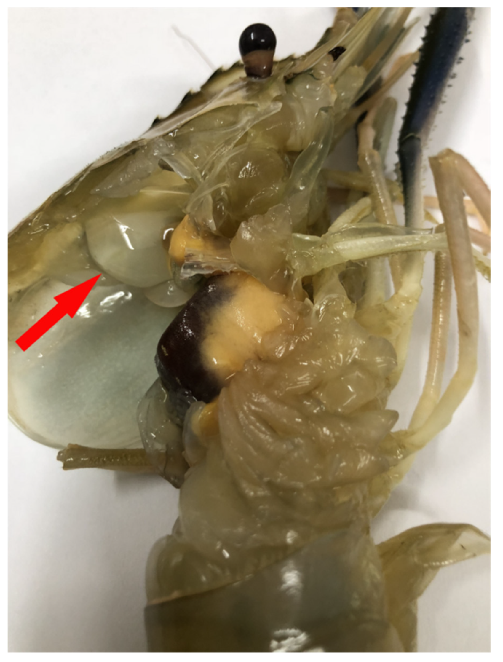
3.1. Clinical Symptoms of WBD in M. rosenbergii

A total of 30 infected M. rosenbergii and 30 healthy M. rosenbergii were used to examine the clinical signs of C. freundii infection. Compared with normal prawns, C. freundii-infected prawns showed a 7 mm-diameter bubble under the carapace of the diseased prawn (Figure 1), loss of appetite, inactivity, and weight loss. The 30 infected prawns all had a water bubble under the carapace. C. freundii infection caused mass mortality of the infected prawns after the onset of clinical symptoms and the rapid spread of the disease between prawns.

3.2. Molecular Identification and Biochemical Characterization

The C. freundii strain GDZQ201912 was a motile, rod shaped, Gram-negative bacterium (Figure 2A). C. freundii was rod-shaped with blunt edges at both ends by TEM (Figure 2B). Ten individuals were randomly measured. The width and length of the bacteria were 0.67–0.76 and 1.88–2.07 µm, respectively.
The 16S rRNA gene amplified from strain GDZQ201912 was sequenced and BLAST analyzed against the non-redundant database, which showed 99.66% identity with C. freundii (GenBank Accession Number: CP060662.1). Table 3 and Table 4 present the physiological and biochemical characteristics of strain GDZQ201912, respectively. The molecular sequence and biochemical analyses revealed that the isolated bacterium was C. freundii.

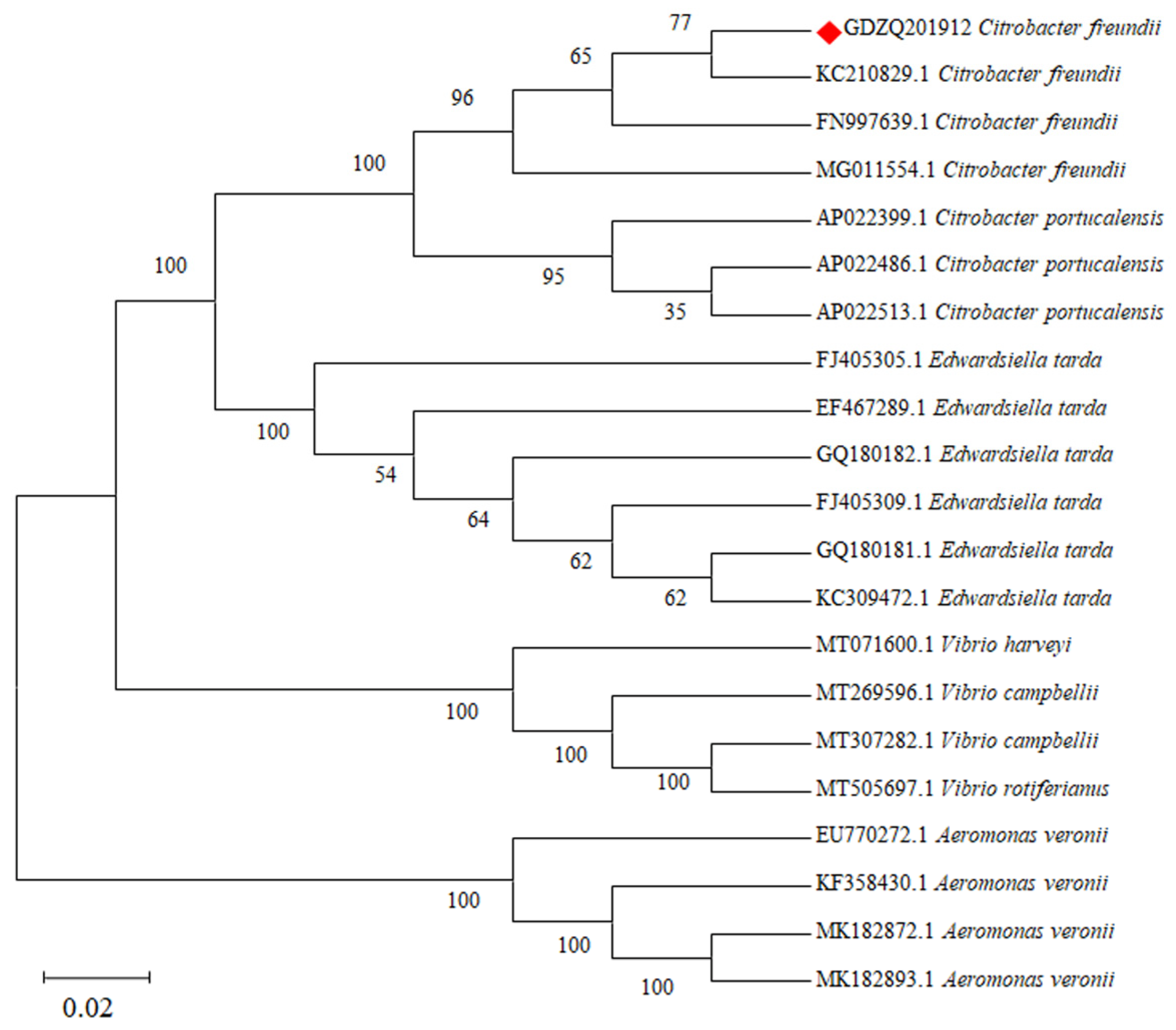
3.3. Phylogenetic Analysis

A phylogenetic tree was constructed using the NJ method by the MEGA 7 software based on the 16S rRNA gene sequences of strain GDZQ201912, and the phylogenetic tree revealed the relationship of strain GDZQ201912 with other Citrobacter and pathogenic bacterium species. Bootstrap values above 50% are shown at the nodes. The 16S rRNA gene sequence of strain GDZQ201912 was found to be evolutionarily close to C. freundii with the highest bootstrap value (Figure 3).

3.4. Effects of Temperature, pH, and Salinity on the Growth of C. freundii

Figure 4 shows the effects of temperature, pH, and salinity on the growth of C. freundii. C. freundii exhibits a wide adaptability to temperature. It can grow well in a range from 24 °C to 42 °C (Figure 4A). The optimum temperature was 32 °C. C. freundii has a wide pH range and can grow normally at pH 4–9, whose optimal value was around pH 6 (Figure 4B). The maximum salt tolerance of C. freundii was 6%, whose optimum value was 1% (Figure 4C). Our results indicated that C. freundii has a wide range of temperature, pH, and salinity tolerance.

3.5. Experimental Challenge

Prawns challenged with strain GDZQ201912 died from the third day post-injection (dpi), and no death occurred in the control groups (Figure 5). The challenged prawns exhibited the typical signs of WBD as described previously. The diseased prawns were similar to those observed in naturally infected prawns in 2019. The bacteria re-isolated from the water bubble of challenged prawns and were reconfirmed as C. freundii.

3.6. LD50 of Strain GDZQ201912 in M. rosenbergii

Figure 6 shows the cumulative mortality rate of M. rosenbergii after post-infection with C. freundii. For each trial, 10 prawns were injected with bacteria inoculum at 100–10−6 dilutions, and the control group was injected with same volume of PBS. The control prawns showed no mortality during the experimental challenge. Among M. rosenbergii injected with serial dilutions of C. freundii, prawns injected with 100, 10−1, and 10−2 dilutions showed disease signs from 2, 3, and 4 dpi, respectively. Prawns injected with 10−3 and 10−4 dilutions exhibited disease symptoms from 5 dpi, whereas no death occurred among those injected with 10−5 and 10−6 dilution during the experimental challenge. The LD50 of C. freundii was 104.94 CFU/g (Figure 6).

3.7. Antibiogram Tests of Strain GDZQ201912

Antibiogram tests of strain GDZQ201912 revealed its resistance to different antibiotics, such as penicillin, ampicillin, clindamycin, lincomycin, and novobiocin, whereas it showed sensitivity to doxycycline, florfenicol, norfloxacin, ofloxacin, amoxicillin, cefepime, ceftriaxone, neomycin, gentamicin, streptomycin, tetracycline, polymyxin B, and rifampin (Table 5). Among the 13 sensitive antibiotics, strain GDZQ201912 was the most sensitive to cefepime, followed ceftriaxone, ofloxacin, and florfenicol in fourth.

3.8. Antibiotic Treatment

The M. rosenbergii mortalities caused by florfenicol and ofloxacin treatment at different time points after the intramuscular challenge with C. freundii or PBS were monitored for 14 days (Figure 7). Prawns fed with common pellets after C. freundii infection died from 5 dpi, and the death reached the maximum on 8 dpi with mortalities of 93.0%. In Group I, prawns fed with florfenicol mixed pellets after C. freundii infection died from 8 dpi, and the death reached the maximum on 8 dpi with mortalities of 19.40%; prawns fed with ofloxacin mixed pellets after C. freundii infection died from 7 dpi, and death reached the maximum on 8 dpi with mortalities of 32.83%. In Group II, prawns fed with florfenicol and ofloxacin mixed pellets after C. freundii infection all died from 5 dpi, and the death reached the maximum on 7 dpi with mortalities of 35.67% and 57.47%, respectively. No death was observed among prawns fed with common pellets and mixed pellets of florfenicol and ofloxacin after PBS injection. The evaluation of the therapeutic effects of antibiotic pellets revealed that the RPS of florfenicol and ofloxacin in group I against C. freundii in prawns were 86.67% and 72.22%, respectively (Figure 7A). The RPS of florfenicol and ofloxacin in Group II against C. freundii in prawns were 69.17% and 47.53%, respectively (Figure 7B). Both florfenicol and ofloxacin initially showed effective protection for M. rosenbergii against C. freundii (and especially florfenicol).

3.9. Immune and Antioxidant Enzyme Activity Changes in Hemolymph

The enzymatic activities of SOD, CAT, GPx, GST, MDA, ACP, ALP, and LZM of M. rosenbergii were measured at 1-, 3-, 5-, 7-, 9-, 11-, and 14-days after the C. freundii strain GDZQ201912 and florfenicol challenge. In hemolymph, the results revealed that the SOD activity in the groups fed with common and florfenicol mixed pellets significantly increased from three days post C. freundii infection, rapidly declined at five and seven days (p < 0.05, Figure 8A), and increased again to normal levels at 9-, 11-, and 14- days compared with the PBS control (fed with common pellets) group (p > 0.05, Figure 8A). No significant differences were observed between the SOD activities in the PBS control (fed with common pellets) groups (p > 0.05, Figure 8A).
The CAT activity in M. rosenbergii fed with common pellets significantly increased from three-, five-, and nine-days post C. freundii infection and reached the peak value at 5 dpi compared with the PBS control (fed with common pellets) group (p < 0.05, Figure 8B). This value significantly reduced to the lowest at 7-dpi. During this period, the CAT activity of prawns fed with florfenicol mixed pellets significantly increased from three and five days post C. freundii infection and reached the peak value at 5-dpi compared with the PBS control (fed with common pellets) group (p < 0.05, Figure 8B). No significant differences were observed between the CAT activities in the PBS control (fed with common pellets) groups (p > 0.05, Figure 8B).
The GPx activity in M. rosenbergii fed with common pellets significantly increased at 3-, 5-, 7-, 9-, 11-, and 14-days post C. freundii infection and reached the peak value at 5-dpi compared with the PBS control (fed with common pellets) group (p < 0.01, Figure 8C). During this period, the groups fed with florfenicol mixed pellets (C. freundii infection) presented similar trend levels of enzymatic activity, which were lower than that observed for the group fed with common pellets (C. freundii infection) and reached the peak value at 5 dpi compared with the PBS control (fed with common pellets) group (p < 0.01, Figure 8C). No significant differences were observed between the GPx activities in the PBS control (fed with common pellets) groups (p > 0.05, Figure 8C).
The GST activity of prawns fed with common mixed pellets significantly increased at 3-, 5-, 7-, 9-, 11-, and 14-days post C. freundii infection and reached the peak value at 3 dpi compared with the PBS control (fed with common pellets) group (p < 0.01, Figure 8D). During this period, the groups fed with florfenicol mixed pellets (C. freundii infection) presented similar trend levels of enzymatic activity, which were lower than that observed for the group fed with common pellets (C. freundii infection) and reached the peak value at 3-dpi compared with the PBS control (fed with common pellets) group (p < 0.01, Figure 8D). No significant differences were observed between the GST activities in the PBS control (fed with common pellets) groups (p > 0.05, Figure 8D).
The MDA activity of prawns fed with common mixed pellets significantly increased at three, five, seven, and nine days post C. freundii infection and reached the peak value at 3-dpi compared with the PBS control (fed with common pellets) group (p < 0.01, Figure 8E). During this period, the groups fed with florfenicol mixed pellets (C. freundii infection) presented similar trend levels of enzymatic activity, which were lower than that observed for the group fed with common pellets (C. freundii infection) and reached the peak value at 3-dpi, compared with the PBS control (fed with common pellets) group (p < 0.05, Figure 8E). No significant differences were observed between the MDA activity in the PBS control (fed with common pellets) groups (p > 0.05, Figure 8E).
The ACP activity in M. rosenbergii fed with common pellets significantly increased from 3-days post C. freundii infection and peaked at 7-dpi (556.34 U/L) compared with the PBS control (fed with common pellets) group (p < 0.01, Figure 8F). Subsequently, this value declined at 9-, 11-, and 14-days. During this period, the two groups fed with florfenicol pellets (PBS and C. freundii infected) presented similar trend levels of enzymatic activity, which were lower than that observed for the group fed with common pellets and reached the peak values of 177.67 and 376.34 U/L on 5- and 7- dpi, respectively (p < 0.01). No significant differences were observed in this parameter in the PBS control (fed with common pellets) group (p > 0.05, Figure 8F).
The ALP activity of prawns fed with common and florfenicol mixed pellets significantly increased at three-, five-, seven-, and nine-days after C. freundii infection, with peak values of 98.67 and 64.67 U/L, respectively, at three and seven days compared with the PBS control (fed with common pellets) group (p < 0.01, Figure 8G). No significant differences were observed between ALP activities in the PBS control (fed with common pellets) group (p > 0.05, Figure 8G).
The LZM activity in M. rosenbergii fed with common pellets significantly increased from 3-, 5-, 7-, 11-, and 14-days post C. freundii infection and peaked 450.00 U/mL at 7 days compared with the PBS control (fed with common pellets) group (p < 0.01, Figure 8H). This value declined to normal level at 9-dpi (p > 0.05, Figure 8H). During this period, the groups fed with florfenicol mixed pellets (C. freundii infection) presented similar trend levels of enzymatic activity, which were lower than that observed for the group fed with common pellets and reached the peak value at seven days (p < 0.05, Figure 8H). No significant differences were observed between the LZM activities in the PBS control (fed with common pellets) groups (p > 0.05, Figure 8H).

3.10. Immune and Antioxidant Enzyme Activity Changes in Hepatopancreas

The results revealed that the SOD activity in M. rosenbergii (fed with common and florfenicol mixed pellets) significantly increased at 3-days, decreased at 5- and 7-days post C. freundii infection (p < 0.05, Figure 9A), and increased again to normal levels at 11 and 14 days compared with the PBS control (fed with common pellets) group (p > 0.05, Figure 9A). No significant differences were observed between the SOD activities in the PBS control (fed with common pellets) groups (p > 0.05, Figure 9A).
The CAT activity in M. rosenbergii (fed with common and florfenicol mixed pellets) significantly increased at three and five days and reached the peak value at 3- and 5-dpi, respectively, while prawns fed with common pellets decreased at seven days post C. freundii injection compared with the PBS control (fed with common pellets) group (p < 0.05, Figure 9B). No significant differences were observed between the CAT activities in the PBS control (fed with common pellets) groups (p > 0.05, Figure 9B).
The GPx activity in M. rosenbergii (fed with common and florfenicol mixed pellets) significantly increased at 3-, 5-, 7-, 9-, and 11-days post C. freundii injection and both reached the peak value at 5-dpi compared with the PBS control (fed with common pellets) group (p < 0.01, Figure 9C). No significant differences were observed between the GPx activities in the PBS control (fed with common pellets) group (p > 0.05, Figure 9C).
The GST activity of prawns (fed with common and florfenicol mixed pellets) significantly decreased at 3-, 5-, 7-, 9-, and 11-days post C. freundii injection, compared with the PBS control (fed with common pellets) group (p < 0.05, Figure 9D). No significant differences were observed between the GST activities in the PBS control (fed with common pellets) and C. freundii infection groups (fed with florfenicol mixed pellets) (p > 0.05, Figure 9D).
The MDA activity of prawns (fed with common and florfenicol mixed pellets) significantly increased at 3-, 5-, and 7-days post C. freundii injection, compared with the PBS control (fed with common pellets) group (p < 0.05, Figure 9E). No significant differences were observed between the MDA activities in the PBS control (fed with common and florfenicol mixed pellets) groups (p > 0.05, Figure 9E).
The ACP activity in the hepatopancreas of M. rosenbergii fed with common and florfenicol mixed pellets significantly increased at 3-, 5-, 7-, 9-, 11-, and 14-days post C. freundii infection and reached the peak value at 5- and 7-dpi, respectively, compared with the PBS control (fed with common pellets) group (p < 0.05, Figure 9F). No significant differences were observed between the ACP activities in the PBS control (fed with common pellets and florfenicol mixed pellets) groups (p > 0.05, Figure 9F).
The ALP activity of prawns (fed with common and florfenicol mixed pellets) significantly increased at three- and five-days post C. freundii injection, compared with the PBS control (fed with common pellets) group (p < 0.05, Figure 9G). No significant differences were observed between the ALP activities in the PBS control (fed with common and florfenicol mixed pellets) groups (p > 0.05, Figure 9G).
The LZM activity in M. rosenbergii fed with common pellets significantly increased at three-, five-, and seven-days post C. freundii infection and reached the peak value at 5-dpi compared with the PBS control (fed with common pellets) group (p < 0.01, Figure 9H). This value declined to normal levels at 9-, 11-, and 14-dpi. During this period, the groups fed with florfenicol mixed pellets (C. freundii infection) presented similar trend levels of enzymatic activity, which were lower than that observed for the prawns fed with common pellets and reached the peak value at 7-days (p < 0.05, Figure 9H). No significant differences were observed between the LZM activities in the PBS control (fed with common and florfenicol mixed pellets) groups (p > 0.05, Figure 9H).

3.11. Expression of Immune-Related Genes in Hepatopancreas after the Challenge with C. freundii

Immunity-related genes (Cu/Zn-SOD, CAT, GPx, GST, LZM, ACP, ALF, crustin, CypA and CTL) mRNA expressions were determined in the hepatopancreas of M. rosenbergii at 1-, 3-, 5-, 7-, 9-, 11-, and 14-days after the challenge with C. freundii (Figure 10). These genes were up-regulated at different levels following the challenge with C. freundii in the groups fed with common and florfenicol mixed pellets compared with the PBS control groups (fed with common and florfenicol mixed pellets). The mRNA expressions of several genes (CAT, GPx, GST, LZM, ACP, ALF, and CypA) in M. rosenbergii fed with common pellets reached their peak at five days after the challenged with C. freundii (Figure 10B–G,I), whereas the mRNA expressions of crustin (Figure 10H) and CTL (Figure 10J) peaked at 3- and 7-dpi, respectively. The mRNA expressions of several genes (CAT, GPx, GST, crustin and CTL) in M. rosenbergii fed with florfenicol mixed pellets reached their peak at 5-dpi (Figure 10B–D,H,J, respectively). Meanwhile, the mRNA expressions of LZM, ACP, ALF and CypA genes peaked at 7-dpi (Figure 10E–G,I, respectively). In addition, the mRNA expression of Cu/ZnSOD in prawns (fed with common and florfenicol mixed pellets) were up-regulated at 3-dpi and down-regulated at 5- and 7-dpi, with the lowest peak value observed at 7-dpi (Figure 10A). No evident difference was reported between the PBS control groups (fed with common pellets).

4. Discussion

Diseases are a major constraint in the sustainable development of aquaculture all over the world. The outbreak and spread of bacterial infections in aquaculture farms pose a major concern. C. freundii is an opportunistic pathogen that is commonly found in water, soil, animals and has been reported to cause numerous diseases in aquaculture [39,40,41]. In recent years, an increasing number of studies reported C. freundii causing diseases and serious mass deaths in various aquatic products, such as Chinese mitten-handed crab [42], red swamp crayfish [14], grass carp [24], and crucian carp [43]. In addition, C. freundii can cause infections in amphibians, reptiles, birds, and mammals [36,44,45,46,47,48]. In the present study, C. freundii isolated from diseased M. rosenbergii was confirmed to be the cause contributing to prawn mortality, with the typical signs of water bubble under the carapace.
Phylogenetic analysis revealed that our isolated strain GDZQ201912 showed a very close association with other strains of C. freundii submitted from China and UK and formed separate clades with other Citrobacter sp., such as C. portucalensis (AP022399.1, AP022486.1, and AP022513.1). The physiological and biochemical profiles of strain GDZQ201912 were similar to those of other isolates reported by earlier works [49,50]. Variations in the biochemical test, such as those of tyrosine, putrescine [38], indole, citrate, H2S, malonate, raffinose, and dulcitol, were also described by earlier studies [17,49].
In the case of pathogenic bacteria, such as C. freundii, multidrug resistance is one of the most prominent problems [50]. In this study, the isolated strain was resistant to four different β-lactam antibiotics, including ampicillin, clindamycin, lincomycin, and penicillin. Numerous studies demonstrated the existence of β-lactamase in C. freundii [51,52,53,54]. The resistant feature against β-lactam antibiotics of our strain indicated the existence of β-lactamase gene, which benefits the strain. The C. freundii resistant to tetracycline and oxytetracycline has been isolated from mammals, poikilothermic hosts, poultry, catfish, and rainbow trout [55,56,57,58,59,60].
The innate immune system defense against invading pathogens relies on AMPs, enzymes, and cellular components [16]. During this process, host cells will produce abundant ROS, such as superoxide anion (O2•−), hydrogen peroxide (H2O2), hydroxyl radicals (OH), and singlet oxygen (1O2), and reactive oxygen intermediates to kill the invading pathogens; however, excessive levels of ROS in the body will also cause the destruction and damage of DNA and other biological macromolecules [18]. Crustaceans can rely on complex enzymatic antioxidant systems to alleviate and restore the oxidative damage, SOD, GPx, GST, CAT, MDA, and so on [61]. SODs conduct the first step in eliminating the oxidative damage caused by ROS from cells. The SOD activity first increased at 1.5 h but decreased significantly at 12–48 h in the hepatopancreas of P. monodon after V. parahaemolyticus infection [62]. In L. vannamei, the SOD activity showed remarkable decreases in the hemolymph after 6–96 h and recovery after 96 h post-V. alginolyticus injection [63]. The SOD activity was also significantly reduced in Probopyrus ringueleti-infected Palaemontes argentinus [64]. In addition, the Cu/ZnSOD showed an up-regulated expression in the hemocytes of M. rosenbergii at 3 and 6 h after Lactococcus garvieae infection, whereas down-regulation was observed in the hepatopancreas at 3 h after L. garvieae infection [65]. In M. nipponense, the ecCuZnSOD mRNA expression level showed a drastic decrease at 12 h after A. hydrophila injection [66]. In M. rosenbergii, the cytMn-SOD transcript also decreased in the hepatopancreas at 3 h after L. garvieae injection [67]. The expression of icCu/Zn-SOD decreased in the first 8–16 h and then recovered after the challenge with Listonella anguillarum and Micrococcus luteus in Chlamys farreri [68]. The relative expression level of icCu/Zn-SOD mRNA increased rapidly at 6 h post infection with Vibrio anguillarum in Venerupis philippinarum, followed by a remarkable decrease, and then increased again [69]. In the present study, a similar expression profile was found in the hepatopancreas of M. rosenbergii after C. freundii infection, in which the relative expression level of Cu/ZnSOD mRNA was up-regulated first and then decrease extremely. Then, the expression level recovered, and SOD activities in hepatopancreas and hemolymph were restored. CAT plays an important role in the antioxidant enzyme defense system to alleviate ROS. The CAT activity in the hepatopancreas of M. rosenbergii and CAT mRNA expression increased markedly at 6 and 24 h post injection with V. parahaemolyticus [70]. The CAT activity exhibited a significant rise at the early stage of white spot syndrome virus infection in P. monodon [71]. The CAT activity in the hepatopancreas of M. rosenbergii significantly decreased at 5 days after spiroplasma MR-1008 infection and showed a recovery at 15 days; however, the mRNA expression level was up-regulated and peaked at 12 days [72]. Similar trends were observed in the CAT activity and mRNA expression level in the present study. GST and GPx are involved in different mechanisms for the host defense against oxidative damage [73]. In Fenneropenaeus chinensis, the mRNA expression levels of FcThetaGST, FcMuGST, and FcGPx in the hepatopancreas were up-regulated after the challenge with V. anguillarum for 24 h, whereas the GST activity decreased [74]. In L. vannamei, the GPx activity and mRNA transcription increased significantly at 12 h after the challenge with V. anguillarum [75]. In P. monodon, the GPx activity of hepatopancreas increased significantly at 6 h and reached the peak level at 12 h after the V. parahaemolyticus challenge; the MDA activity significantly increased from 6 h to 24 h with a peak at 6 h, whereas the GST activity decreased significantly at 3 h [62]. The GPx activity in the gill of F. chinensis increased at 6 h and remained at high levels up to 24 h [74]. In addition, the results of increased mRNA expression levels and activities (GPx, GST, CAT, and MDA) during C. freundii infection in M. rosenbergii were in agreement with the histopathological damage in hepatopancreas.
LZM is an important enzyme of the innate immune defense system against bacterial infection, and it can cause bacterial cell disintegration by hydrolyzing the bacterial cell wall [76,77]. Lysosomal enzymes, which are produced by phagocytes from crayfish Procambarus clarkii, can efficiently degrade and eliminate foreign materials, as had been demonstrated by Franchini and Ottaviani [78]. In P. monodon and M. rosenbergii, the lysosomal activity showed a significant increase after V. vulnificus infection and increased in L. vannamei infected with Micrococcus lysodeikticus [79,80]. ACP and ALP are symbols of macrophage activation, important components of the lysosome system, and play important roles in the innate immune system to engulf antigens intracellularly [21]. The ACP activity increased substantially within 48 h in the hemolymph of C. farreri after the treatment with V. anguillarum [81]. In L. vannamei, the ACP and ALP activities significantly increased with V. parahaemolyticus and M. lysodeikticus infections, respectively [82]. In addition, the ACP and ALP activities in M. rosenbergii were significantly enhanced after the challenge with a novel pathogen spiroplasma MR-1008 [72]. In the present study, the enzyme activities of ACP, ALP, and LZM in the hepatopancreas and hemolymph were all enhanced after M. rosenbergii infection with C. freundii; the enzyme activities all increased significantly in the hepatopancreas after T. chinensis infection in M. nipponense [83].
AMPs are the main effector molecules in the innate immunity of crustaceans, and they act as frontline effectors to defend against invading bacteria, fungi, and viruses [26,27,84]. The major AMPs, such as penaeidins, crustins, and ALF, have been identified in shrimp [29]. In P. monodon, the mRNA expression level of crustin-like peptide significantly increased at 24 h post-infection with V. harveyi and recovered at 72 h [85]. The mortality of crustin-depleted L. vannamei by RNA interference (RNAi) had a remarkable increase within 48 h post-infection with V. penaeicida [86]. In Marsupenaeus japonicus, MjCru I-1 can enhance the hemocyte phagocytosis, increase shrimp survival rate with V. anguillarum and Staphylococcus aureus infection, and weaken the bacterial clearance by knockdown of MjCru I-1 [84]. In L. vannamei, LvCrustinB was up-regulated with V. parahaemolyticus infection, and the knockdown of LvCrustinB can increase the shrimp mortality rate [87]. The expression level of FcALF2 in Fenneropenaeus chinensis significantly increased after the injection with M. lysodeikticus and V. anguillarum [88]. The in vivo function of LvALF1 in response to V. penaeicida and Fusarium oxysporum infections was measured by RNAi; the LvALF1-depleted L. vannamei caused a significant increase in the mortality of infected shrimps [89]. With C. freundii infection, the ALF mRNA expression significantly increased in crayfish P. clarkii [14]. The expressions of ALF and crustin increased in the hepatopancreas of M. rosenbergii after C. freundii infection in the present study; in addition, the ALF expression significantly increased in L. vannamei after the V. anguillarum and M. lysodeikticus challenge [90].
CypA acts as the receptor for the immunosuppressive agent cyclosporin A and belongs to the superfamily of peptidyl-prolyl cis-trans isomerases [91]. The mRNA expression of btsCypA was up-regulated in the hepatopancreas of P. monodon after stimulation by lipopolysaccharide [92]. After V. anguillarum challenge, the expression level of CfCypA was up-regulated in the gonads of C. farreri and reached the peak at 4 h post-injection [93]. The mRNA expression level of EsCypA increased in the fungal Pichia pastoris-infected E. sinensis [24]. The significantly up-regulated VpCypA2 mRNA expression level was observed in the hemocytes of V. philippinarum after the challenge with L. anguillarum [94]. The expression of CypA increased in the hepatopancreas of M. rosenbergii after C. freundii infection in the present study; the CypA mRNA expression level was significantly up-regulated in the hepatopancreas of P. clarkii after the challenge with C. freundii [14].
CTLs are key recognition proteins and play important roles in shrimp innate immunity for the recognition and clearance of pathogens [22,23,24,25]. PmCL1 expression was significantly upregulated in the hepatopancreas and gill of P. monodon after the infection with V. harveyi and V. anguillarum, respectively [95]. The expressions of hepatopancreas-specific CTL (Fc-hsL) and CTLs (Fclectin, FcLec3 and FcLec4) from F. chinensis were up-regulated following the challenge of shrimp with V. anguillarum or S. aureus [96,97,98,99]. In L. vannamei, the expressions of LvCTL3 and LvPLP (the perlucin-like protein, a typical CTL) were up-regulated after V. parahaemolyticus infection; two CTLs (LvLectin-1 and LvLectin-2) showed significantly increased expressions after the L. anguillarum challenge [100,101,102]. Two novel lectins (MnCTLDcp2 and MnCTLDcp3) from M. nipponense were up-regulated in the heart after the challenge with A. hydrophila [103]. The expression of CTL increased in the hepatopancreas of M. rosenbergii after C. freundii infection in the present study, and a significantly up-regulated CTL mRNA expression level was observed in the hepatopancreas of P. clarkii after the challenge with C. freundii [14].
In general, when M. rosenbergii was infected with C. freundii, CTL, as one of pattern-recognition receptor, immediately recognized the non-self material that had entered the prawn body. With C. freundii infection, enzymes involved in the antioxidant antibacterial system (SOD, CAT, GPx, GST, and MDA) were synthesized rapidly in the hepatopancreas and hemolymph, and the phosphatase enzymes (ACP and ALP) and LZM also actively participated in this immune response. In addition, the expressions of immune-related genes (Cu/Zn-SOD, CAT, GPx, GST, LZM, ACP, ALF, crustin, CypA, and CTL) in the hepatopancreas were significantly up- or down- regulated, which suggested that the immediate immune response system in M. rosenbergii offers defense against C. freundii infection. The results of this study indicated that all of these antioxidant enzymes and genes play important roles in the M. rosenbergii immune responses. Furthermore, the hepatopancreas are important immune tissues in the defense against C. freundii.
Antibiotics are natural or synthetic compounds and have become the main means for the prevention and treatment of infectious diseases due to their low cost, good curative effect, and simple operation [104,105]. Florfenicol, oxytetracycline, oxytetracycline hydrochloride, ormetoprim, and sulfamethazine were the five antibiotics approved by the FDA [106]. In the present study, florfenicol and ofloxacin were selected to evaluate their therapeutic effects against C. freundii in M. rosenbergii. Ofloxacin was forbidden in aquaculture and its use in the present study was just for scientific research. Florfenicol is a broad-spectrum and effective antibacterial, belonging to a part of the chloramphenicol family of drugs, that is widely used to control susceptible bacterial diseases in fish and shrimp farming [107,108,109,110,111]. Florfenicol and ofloxacin can stop bacteria from multiplying by interfering with bacterial protein production and DNA replication, respectively [31]. The increasd immune and antioxidant responses in M. rosenbergii after C. freundii challenge were results of C. freundii activated the host’s innate immune system. With antibiotics feeding, C. freundii in M. rosenbergii cannot multiplying well owing to the protein production or DNA replication interfering. Then, the innate immune system of C. freundii infected M. rosenbergii was more efficient against bacteria with the higher survival rate, immune and antioxidant responses. C. freundii can break the innate immune system of M. rosenbergii treated without antibiotics, damage the capital functional organs and cause death. Florfenicol was effective in the treatment of pseudotuberculosis in Yellowtail Seriola quinqueradiata [112], vibriosis in Goldfish Carassius auratus [113], and furunculosis in Atlantic Salmon Salmo salar [114], V. anguillarum infection in Atlantic cod Gadus morhua [106], vibriosis in Black Tiger Shrimps P. monodon [111], V. harveyi infection in L. vannamei [110]. Due to the adverse environmental consequences, alternatives to antibiotics have been developed, including probiotics, phage therapy, essential oils (EOs), and Chinese herbal medicine [31]. Pseudomonas synxantha and Pseudomonas aeruginosa-fed Penaeus latisulcatus showed a higher tolerance to the V. harveyi challenge [115]. Four phages that had lytic activity against V. harveyi were isolated, and were demonstrated to be effective in controlling the population of V. harveyi in hatchery systems and improved the survival of P. monodon [116]. The usage of EOs from Cinnamosma fragrans enhanced the survival of P. monodon larvae with Vibrio penaeicidae and Vibrio splendidus challenge and decreased bacterial concentration [117]. For the WBD in M. rosenbergii, we also conducted alternatives therapies, such as Chinese herbal medicine. We found strain GDZQ201912 was sensitive to Terminalia chebula Retz, Scutellaria baicalensis Georgi, Caesalpinia sappan Linn, Rhus chinensis Mill, and Schisandra chinensis as well as the evaluations of their therapies of WBD, survival rate, immune and antioxidant responses of M. rosenbergii (Unpublished data).

5. Conclusions

Overall, in the present study, the pathogenic bacterium C. freundii was isolated from WBD M. rosenbergii with a mass mortality, and the cause of WBD in M. rosenbergii was identified with the typical sign under the carapace. The TEM observation, physiological and biochemical characteristics, LD50, and antimicrobial susceptibility of the C. freundii isolate were studied. The therapeutic effects of antibiotics against WBD were also evaluated, and florfenicol was the most efficient antibiotic to defend against C. freundii. In addition, the results of the present study provide insights into the histopathological changes, antioxidant enzymatic activity, and changes in the expressions of immune-related genes of M. rosenbergii challenged with C. freundii, revealing the antibacterial immune responses in giant river prawn. These results will facilitate the development of therapies and management of the disease caused by this pathogen in aquaculture systems.

Author Contributions

Conceptualization, J.H.; methodology, C.Z.; software, H.W.; validation, C.Z., H.W. and S.H.; formal analysis, C.Z.; investigation, S.W.; resources, J.H.; data curation, C.Z.; writing—original draft preparation, C.Z.; writing—review and editing, J.H.; visualization, C.Z.; supervision, J.H. and C.Z.; project administration, S.W.; funding acquisition, J.H. All authors have read and agreed to the published version of the manuscript.

Funding

This work was funded by the National Key Research and Development Program of China (2018YFD0900501), Key-Area Research and Development Program of Guangdong Province (2019B020217001) and Supported by the earmarked fund for CARS-48.

Institutional Review Board Statement

Not applicable.

Informed Consent Statement

Not applicable.

Data Availability Statement

The data presented in this study are available in the article.

Conflicts of Interest

The authors declare no conflict of interest.

References

  1. Nhan, D.T.; Wille, M.; Sorgeloos, P. Comparison of reproductive performance and offspring quality of giant freshwater prawn (Macrobrachium rosenbergii) broodstock from different regions. Aquaculture 2009, 298, 36–42. [Google Scholar] [CrossRef]
  2. New, M. Freshwater prawn farming: Global status, recent research and a glance at the future. Aquac. Res. 2005, 36, 210–230. [Google Scholar] [CrossRef]
  3. Kumaresan, V.; Palanisamy, R.; Pasupuleti, M.; Arockiaraj, J. Impacts of environmental and biological stressors on immune system of Macrobrachium rosenbergii. Rev. Aquacult. 2017, 9, 283–307. [Google Scholar] [CrossRef]
  4. FAO Data form “Fisheries and Aquaculture Software Fishstatj-Software for Fishery and Aquaculture Statistical Time Series”. FAO Fisheries Division 2021, Rome, Italy. Available online: http://www.fao.org/fishery/statistics/software/fishstatj/en (accessed on 18 November 2021).
  5. Tiruvayipati, S.; Bhassu, S. Host, pathogen and the environment: The case of Macrobrachium rosenbergii, Vibrio parahaemolyticus and magnesium. Gut. Pathog. 2016, 8, 15. [Google Scholar] [CrossRef] [PubMed] [Green Version]
  6. Gao, X.J.; Miao, Z.; Li, X.X.; Chen, N.; Gu, W.W.; Liu, X.D.; Yang, H.; Wei, W.H.; Zhang, X.J. Pathogenicity of non-O1/O139 Vibrio cholerae and its induced immune response in Macrobrachium rosenbergii. Fish. Shellfish. Immunol. 2019, 92, 300–307. [Google Scholar] [CrossRef] [PubMed]
  7. Zhang, Y.Y.; Wang, L.F.; Zhuang, H.; Li, X.X.; Gao, X.J.; An, Z.H.; Liu, X.D.; Yang, H.; Wei, W.Z.; Zhang, X.J. Excessive use of enrofloxacin leads to growth inhibition of juvenile giant freshwater prawn Macrobrachium rosenbergii. Ecotoxicol. Environ. Saf. 2019, 169, 344–352. [Google Scholar] [CrossRef] [PubMed]
  8. Zhu, X.J.; Yang, X.Q.; He, W.R.; Xiong, Y.A.; Liu, J.; Dai, Z.M. Involvement of tetraspanin 8 in the innate immune response of the giant prawn, Macrobrachium rosenbergii. Fish. Shellfish. Immunol. 2019, 86, 459–464. [Google Scholar] [CrossRef] [PubMed]
  9. Tao, B.H.; Shi, H.R.; Huang, J.W.; Wang, G. Studied on yellow and black gills of Macrobrachium rosenbergii caused by Pseudoonas. Acta Sci. Nat. Univ. Sunyatseni 2000, 1, 255–259. [Google Scholar]
  10. Cheng, W.; Chen, J. Isolation and characterization of an Enterococcus-like bacterium causing muscle necrosis and mortality in Macrobrachium rosenbergii in Taiwan. Dis. Aquat. Organ. 1998, 34, 93. [Google Scholar] [CrossRef] [PubMed] [Green Version]
  11. Ramalingam, K.; Ramarani, S. Effect of Pseudomonas aeruginosa on the giant freshwater prawn, Macrobrachium rosenbergii histopathological and electron microscopic study. J. Environ. Biol. 2007, 28, 627–635. [Google Scholar] [PubMed]
  12. Jayaprakash, N.S.; Rejish, K.V.J.; Philip, R.; Bright, S.I.S. Vibrios associated with Macrobrachium rosenbergii (De Man, 1879) larvae from three hatcheries on the Indian southwest coast. Aquac. Res. 2006, 37, 351–358. [Google Scholar] [CrossRef]
  13. Sung, H.H.; Hwang, S.F.; Tasi, F.M. Responses of giant freshwater prawn (Macrobrachium rosenbergii) to challenge by two strains of Aeromonas spp. J. Invertebr. Pathol. 2000, 76, 278–284. [Google Scholar] [CrossRef] [PubMed]
  14. Liu, X.; He, X.; An, Z.; Sun, W.; Chen, N.; Gao, X.; Li, X.X.; Zhang, X. Citrobacter freundii infection in red swamp crayfish (Procambarus clarkii) and host immune-related gene expression profiles. Aquaculture 2020, 515, 734499. [Google Scholar] [CrossRef]
  15. Li, Y.; Zhan, F.; Li, F.; Lu, Z.; Shi, F.; Xu, Z.; Yang, Y.C.; Zhao, L.J.; Qin, Z.D.; Lin, L. Immune function of cytosolic manganese superoxide dismutase from Macrobrachium rosenbergii in response to bacterial infection. Aquaculture 2021, 541, 736771. [Google Scholar] [CrossRef]
  16. Butprom, S.; Phumkhachorn, P.; Rattanachaikunsopon, P. Effect of Lactobacillus plantarum C014 on innate immune response and disease resistance against Aeromonas hydrophila in hybrid catfish. Sci. World. J. 2013, 2013, 392523. [Google Scholar] [CrossRef] [PubMed] [Green Version]
  17. Behera, B.K.; Paria, P.; Das, A.; Das, B.K. Molecular identification and pathogenicity study of virulent Citrobacter freundii associated with mortality of farmed Labeo rohita (Hamilton 1822), in India. Aquaculture 2022, 547, 737437. [Google Scholar] [CrossRef]
  18. Li, Y.; Yan, L.; Kong, X.; Chen, J.; Zhang, H. Cloning, expression, and characterization of a novel superoxide dismutase from deep-sea sea cucumber. Int. J. Bio. Macromol. 2020, 163, 1875–1883. [Google Scholar] [CrossRef] [PubMed]
  19. Cetinkale, O.; Belce, A.; Konukoglu, D.; Senyuva, C.; Gumustas, M.K.; Tas, T. Evaluation of lipid peroxidation and total antioxidant status in plasma of rats following thermal injury. Burns 1997, 23, 114–116. [Google Scholar] [CrossRef]
  20. Lopes, P.A.; Pinheiro, T.; Santos, M.C.; da Luz Mathias, M.; Collares-Pereira, M.J.; Viegas-Crespo, A.M. Response of antioxidant enzymes in freshwater fish populations (Leuciscus alburnoides complex) to inorganic pollutants exposure. Sci. Total Environ. 2001, 280, 153–163. [Google Scholar] [CrossRef]
  21. Yin, F.; Gong, H.; Ke, Q.; Li, A. Stress, antioxidant defence and mucosal immune responses of the large yellow croaker Pseudosciaena crocea challenged with Cryptocaryon irritans. Fish. Shellfish. Immunol. 2015, 47, 344–351. [Google Scholar] [CrossRef] [PubMed]
  22. Zhang, X.W.; Xu, W.T.; Wang, X.W.; Mu, Y.; Zhao, X.F.; Yu, X.Q.; Wang, J.X. A novel C-type lectin with two CRD domains from Chinese shrimp Fenneropenaeus chinensis functions as a pattern recognition protein. Mol. Immunol. 2009, 46, 1626–1637. [Google Scholar] [CrossRef] [PubMed]
  23. Zhang, Y.X.; Zhang, M.L.; Wang, X.W. C-Type Lectin Maintains the Homeostasis of Intestinal Microbiota and Mediates Biofilm Formation by Intestinal Bacteria in Shrimp. J. Immunol. 2021, 206, 1140–1150. [Google Scholar] [CrossRef] [PubMed]
  24. Wang, M.; Zhang, D.; Wang, L.; Gai, Y.; Zhou, Z.; Zhang, H.; Song, L. The molecular characterization of a cyclophilin A from Chinese mitten crab Eriocheir sinensis and the antifungal activity of its recombinant protein. Electron. J. Biotechn. 2013, 16, 2. [Google Scholar] [CrossRef]
  25. Wang, X.W.; Vasta, G.R.; Wang, J.X. The functional relevance of shrimp C-type lectins in host-pathogen interactions. Dev. Comp. Immunol. 2020, 109, 103708. [Google Scholar] [CrossRef] [PubMed]
  26. Brown, K.L.; Hancock, R.E. Cationic host defense (antimicrobial) peptides. Curr. Opin. Immunol. 2006, 18, 24–30. [Google Scholar] [CrossRef] [PubMed]
  27. Hancock, R.E.; Brown, K.L.; Mookherjee, N. Host defence peptides from invertebrates-emerging antimicrobial strategies. Immunobiology 2006, 211, 315–322. [Google Scholar] [CrossRef]
  28. Tassanakajon, A.; Amparyup, P.; Somboonwiwat, K.; Supungul, P. Cationic antimicrobial peptides in penaeid shrimp. Mar. Biotechnol. 2011, 13, 639–657. [Google Scholar] [CrossRef] [PubMed]
  29. Destoumieux, D.; Munoz, M.; Bulet, P.; Bachere, E. Penaeidins, a family of antimicrobial peptides from penaeid shrimp (Crustacea, Decapoda). Cell. Mol. Life. Sci. 2000, 57, 1260–1271. [Google Scholar] [CrossRef]
  30. Liu, B.; Ge, X.; He, Y.; Xie, J.; Xu, P.; He, Y.; Zhaou, Q.; Pan, L.; Chen, R. Effects of anthraquinones extracted from Rheum officinale Bail on the growth, non-specific immune response of Macrobrachium rosenbergii. Aquaculture 2010, 310, 13–19. [Google Scholar] [CrossRef]
  31. Romero, J.; Feijoó, C.G.; Navarrete, P. Antibiotics in aquaculture-use, abuse and alternatives. HEV. Aquacult. 2012, 159, 159–198. [Google Scholar]
  32. Lee, I.M.; Hammond, R.W.; Davis, R.E.; Gundersen, D.E. Universal amplification and analysis of pathogen 16S rDNA for classification and identification of mycoplasmalike organisms. Phytopathology 1993, 83, 834–842. [Google Scholar] [CrossRef]
  33. Lalitha, M.K. Manual on antimicrobial susceptibility testing. In Performance Standards for Antimicrobial Testing: Twelfth Informational Supplement; Clinical and Laboratory Standards Institute: Wayne, PA, USA, 2004; Volume 56238, pp. 454–456. [Google Scholar]
  34. Wayne, P.A. Clinical and laboratory standards institute. Performance standards for antimicrobial susceptibility testing. Inf. Suppl. 2011, 31, 100–121. [Google Scholar]
  35. Kärber, G. Beitrag zur kollektiven Behandlung pharmakologischer Reihenversuche. Arch. Exptl. Pathol. Pharmakol. 1931, 162, 480–483. [Google Scholar] [CrossRef]
  36. Pan, L.; Yang, Y.; Peng, Y.; Li, D.; Khan, T.A.; Chen, P.; Yan, P.; Hu, S.B.; Ding, X.Z.; Sun, Y.J.; et al. The novel pathogenic Citrobacter freundii (CFC202) isolated from diseased crucian carp (Carassius auratus) and its ghost vaccine as a new prophylactic strategy against infection. Aquaculture 2021, 533, 736190. [Google Scholar] [CrossRef]
  37. Chen, M.; Chen, X.Q.; Tian, L.X.; Liu, Y.J.; Niu, J. Improvement of growth, intestinal short-chain fatty acids, non-specific immunity and ammonia resistance in Pacific white shrimp (Litopenaeus vannamei) fed dietary water-soluble chitosan and mixed probiotics. Comp. Biochem. Physiol. C Toxicol. Pharmacol. 2020, 236, 108791. [Google Scholar] [CrossRef]
  38. Frederiksen, W. Citrobacter. In Bergey’s Manual of Systematics of Archaea and Bacteria; Wiley: Hoboken, NJ, USA, 2015; pp. 1–23. [Google Scholar]
  39. Baldissera, M.D.; Souza, C.F.; Junior, G.B.; Moreira, K.L.S.; da Veiga, M.L.; da Rocha, M.; Baldisserotto, B. Citrobacter freundii impairs the phosphoryl transfer network in the gills of Rhamdia quelen: Impairment of bioenergetics homeostasis. Microb. Pathog. 2018, 117, 157–161. [Google Scholar] [CrossRef] [PubMed]
  40. Lü, A.; Hu, X.; Xue, J.; Zhu, J.; Wang, Y.; Zhou, G. Gene expression profiling in the skin of zebrafish infected with Citrobacter freundii. Fish. Shellfish. Immunol. 2012, 32, 273–283. [Google Scholar] [CrossRef]
  41. Sun, H.Y.; Cao, X.H.; Jiang, Y.F.; Ni, L.Y.; Mo, Z.Q.; Qin, Q.W.; Li, Y.W.; Dan, X.M. Outbreak of a novel disease associated with Citrobacter freundii infection in freshwater cultured stingray, Potamotrygon motoro. Aquaculture 2018, 492, 35–39. [Google Scholar] [CrossRef]
  42. Huang, X.D.; Zhou, H.H.; An, J.; Yang, X.L.; Cao, H.P. Isolation, identification and antibiotic susceptibility of pathogenic Citrobacter freundii from Eriocheir sinensis. J. South. Agric. 2019, 50, 1613–1619. [Google Scholar]
  43. Xiong, F.; Qin, L.; Hao, Y.T.; Zhao, D.; Li, W.X.; Zou, H.; Li, M.; Wu, S.G.; Wang, G.T. Gut microbiota modulation and immunity response induced by Citrobacter freundii strain GC01 in grass carp (Ctenopharyngodon idellus). Aquaculture 2020, 521, 735015. [Google Scholar] [CrossRef]
  44. Mauel, M.J.; Miller, D.L.; Frazier, K.S.; Hines, M.E. Bacterial pathogens isolated from cultured bullfrogs (Rana Castesbeiana). J. Vet. Diag. Inv. 2002, 14, 431–433. [Google Scholar] [CrossRef] [PubMed] [Green Version]
  45. Novak, S.S.; Seigel, R.A. Gram-negative septicemia in American alligators (Alligator mississippiensis). J. Wildl. Dis. 1986, 22, 484–487. [Google Scholar] [CrossRef] [PubMed] [Green Version]
  46. Bover-Cid, S.; Holzapfel, W.H. Improved screening procedure for biogenic amine production by lactic acid bacteria. Int. J. Food. Microbiol. 1999, 53, 33–41. [Google Scholar] [CrossRef]
  47. Chuang, Y.M.; Tseng, S.P.; Teng, L.J.; Ho, Y.C.; Hsueh, P.R. Emergence of cefotaxime resistance in Citrobacter freundii causing necrotizing fasciitis and osteomyelitis. J. Inf. 2006, 53, e161–e163. [Google Scholar] [CrossRef] [PubMed]
  48. Brock, T.D. Robert Koch: A Life in Medicine and Bacteriology; Zondervan: Grand Rapids, MI, USA, 1999. [Google Scholar]
  49. Lü, A.; Hu, X.; Zheng, L.; Zhu, A.; Cao, C.; Jiang, J. Isolation and characterization of Citrobacter spp. from the intestine of grass carp Ctenopharyngodon idellus. Aquaculture 2011, 313, 156–160. [Google Scholar] [CrossRef]
  50. Kumar, P.; Meghvansi, M.K.; Kamboj, D.V. Phenotypic characterization and whole-genome analysis of a novel bacteriophage HCF1 infecting Citrobacter amalonaticus and C. freundii. Front. Microbiol. 2021, 12, 15. [Google Scholar] [CrossRef] [PubMed]
  51. Tajima, M.; Takenouchi, Y.; Sugawara, S.; Inoue, M.; Mitsuhashi, S. Purification and properties of chromosomally mediated β-lactamase from Citrobacter freundii GN7391. Microbiology 1980, 121, 449–456. [Google Scholar] [CrossRef] [PubMed] [Green Version]
  52. Almeida, M.V.A.D.; Cangussú, Í.M.; Carvalho, A.L.S.D.; Brito, I.L.P.; Costa, R.A. Drug resistance, AmpC-β-lactamase and extended-spectrum β-lactamase-producing Enterobacteriaceae isolated from fish and shrimp. Rev. Inst. Med. Trop. 2017, 59, e70. [Google Scholar] [CrossRef] [PubMed]
  53. Chen, C.M.; Huang, M.; Wu, H.J.; Guo, M.K.; Wu, L.T. Identification of CFE-2, a new plasmid-encoded AmpC β-lactamase from a clinical isolate of Citrobacter freundii. Int. J. Antimicrob. Ag. 2018, 52, 421–424. [Google Scholar] [CrossRef] [PubMed]
  54. Hasan, M.S.; Sultana, M.; Hossain, M.A. Complete genome arrangement revealed the emergence of a poultry origin superbug Citrobacter portucalensis strain NR-12. J. Glob. Antimicrob. Resist. 2019, 18, 126–129. [Google Scholar] [CrossRef] [PubMed]
  55. Toranzo, A.E.; Cutrin, J.M.; Roberson, B.S.; Nunez, S.; Abell, J.M.; Hetrick, F.M.; Baya, A.M. Comparison of the taxonomy, serology, drug resistance transfer, and virulence of Citrobacter freundii strains from mammals and poikilothermic hosts. App. Eenviron. Microb. 1994, 60, 1789–1797. [Google Scholar] [CrossRef] [PubMed] [Green Version]
  56. Duman, M.; Saticioglu, I.B.; Buyukekiz, A.G.; Balta, F.; Altun, S. Molecular characterization and antimicrobial resistance profile of atypical Citrobacter gillenii and Citrobacter sp. isolated from diseased rainbow trout (Oncorhynchus mykiss). J. Glob. Antimicrob. Resist. 2017, 10, 136–142. [Google Scholar] [CrossRef] [PubMed]
  57. Ture, M.; Kutlu, I. Isolation of Citrobacter freundii from Rainbow Trout (Oncorhynchus mykiss) in Freshwater Cage. J. Limnol. Freshw. Fish. Res. 2018, 4, 85–89. [Google Scholar] [CrossRef]
  58. Zhao, C.Y.; Fu, H.T.; Sun, S.M.; Qiao, H.; Zhang, W.Y.; Jin, S.B.; Gong, Y.S. Experimental inoculation of oriental river prawn Macrobrachium nipponense with white spot syndrome virus (WSSV). Dis. Aquat. Organ. 2017, 126, 125–134. [Google Scholar] [CrossRef]
  59. Jiravanichpaisal, P.; Roos, S.; Edsman, L.; Liu, H.; Söderhäll, K. A highly virulent pathogen, Aeromonas hydrophila, from the freshwater crayfish Pacifastacus leniusculus. J. Invertebr. Pathol. 2009, 101, 56–66. [Google Scholar] [CrossRef] [PubMed]
  60. Khimmakthong, U.; Sukkarun, P. The spread of Vibrio parahaemolyticus in tissues of the Pacific white shrimp Litopenaeus vannamei analyzed by PCR and histopathology. Microb. Pathog. 2017, 113, 107–112. [Google Scholar] [CrossRef] [PubMed]
  61. Jiang, Q.; Jiang, Z.; Ao, S.; Gao, X.; Zhu, X.; Zhang, Z.; Zhang, X. Multi-biomarker assessment in the giant freshwater prawn Macrobrachium rosenbergii after deltamethrin exposure. Ecotoxicol. Environ. Saf. 2021, 214, 112067. [Google Scholar] [CrossRef] [PubMed]
  62. Duan, Y.; Zhang, J.; Dong, H.; Wang, Y.; Liu, Q.; Li, H. Oxidative stress response of the black tiger shrimp Penaeus monodon to Vibrio parahaemolyticus challenge. Fish. Shellfish. Immunol. 2015, 46, 354–365. [Google Scholar] [CrossRef] [PubMed]
  63. Li, C.C.; Yeh, S.T.; Chen, J.C. The immune response of white shrimp Litopenaeus vannamei following Vibrio alginolyticus injection. Fish. Shellfish. Immunol. 2008, 25, 853–860. [Google Scholar] [CrossRef]
  64. Neves, C.A.; Santos, E.A.; Bainy, A.C.D. Reduced superoxide dismutase activity in Palaemonetes argentinus (Decapoda, Palemonidae) infected by Probopyrus ringueleti (Isopoda, Bopyridae). Dis. Aquat. Organ. 2000, 39, 155–158. [Google Scholar] [CrossRef] [Green Version]
  65. Cheng, W.; Tung, Y.H.; Liu, C.H.; Chen, J.C. Molecular cloning and characterisation of copper/zinc superoxide dismutase (Cu, Zn-SOD) from the giant freshwater prawn Macrobrachium rosenbergii. Fish. Shellfish. Immunol. 2006, 21, 102–112. [Google Scholar] [CrossRef]
  66. Xiu, Y.; Wu, T.; Du, J.; Yao, W.; Li, W.; Ding, Z.; Ren, Q.; Gu, W.; Meng, Q.; Wang, W. Molecular characterization and expression analysis of extracellular copper/zinc superoxide dismutase (ecCuZnSOD) from oriental river prawn, Macrobrachium nipponense. Aquaculture 2013, 380, 23–28. [Google Scholar] [CrossRef]
  67. Cheng, W.; Tung, Y.H.; Liu, C.H.; Chen, J.C. Molecular cloning and characterisation of cytosolic manganese superoxide dismutase (cytMn-SOD) from the giant freshwater prawn Macrobrachium rosenbergii. Fish. Shellfish. Immunol. 2006, 20, 438–449. [Google Scholar] [CrossRef] [PubMed]
  68. Ni, D.; Song, L.; Gao, Q.; Wu, L.; Yu, Y.; Zhao, J.; Qiu, L.; Zhang, H.; Shi, F. The cDNA cloning and mRNA expression of cytoplasmic Cu, Zn superoxide dismutase (SOD) gene in scallop Chlamys farreri. Fish. Shellfish. Immunol. 2007, 23, 1032–1042. [Google Scholar] [CrossRef] [PubMed]
  69. Li, C.; Sun, H.; Chen, A.; Ning, X.; Wu, H.; Qin, S.; Xue, Q.; Zhao, J. Identification and characterization of an intracellular Cu, Zn-superoxide dismutase (icCu/Zn-SOD) gene from clam Venerupis philippinarum. Fish. Shellfish. Immunol. 2010, 28, 499–503. [Google Scholar] [CrossRef] [PubMed]
  70. Du, J.; Zhu, H.; Ye, M.; Ma, Y. Macrobrachium rosenbergii Cu/Zn superoxide dismutase (Cu/Zn SOD) expressed in Saccharomyces cerevisiae and evaluation of the immune function to Vibrio parahaemolyticus. Fish. Shellfish. Immunol. 2019, 90, 363–375. [Google Scholar] [CrossRef]
  71. Mathew, S.; Kumar, K.A.; Anandan, R.; Nair, P.G.V.; Devadasan, K. Changes in tissue defence system in white spot syndrome virus (WSSV) infected Penaeus monodon. Comp. Biochem. Physiol. C Toxicol. Pharmacol. 2007, 145, 315–320. [Google Scholar] [CrossRef] [PubMed]
  72. Du, J.; Zhu, H.; Liu, P.; Chen, J.; Xiu, Y.; Yao, W.; Wu, T.; Ren, Q.; Meng, Q.; Gu, W.; et al. Immune responses and gene expression in hepatopancreas from Macrobrachium rosenbergii challenged by a novel pathogen spiroplasma MR-1008. Fish. Shellfish. Immunol. 2013, 34, 315–323. [Google Scholar] [CrossRef] [PubMed]
  73. Kim, M.; Ahn, I.Y.; Cheon, J.; Park, H. Molecular cloning and thermal stress-induced expression of a pi-class glutathione S-transferase (GST) in the Antarctic bivalve Laternula elliptica. Comp. Biochem. Physiol. Part A Mol. Integr. Physiol. 2009, 152, 207–213. [Google Scholar] [CrossRef] [PubMed]
  74. Ren, Q.; Sun, R.R.; Zhao, X.F.; Wang, J.X. A selenium-dependent glutathione peroxidase (Se-GPx) and two glutathione S-transferases (GSTs) from Chinese shrimp (Fenneropenaeus chinensis). Comp. Biochem. Physiol. C Toxicol. Pharmacol. 2009, 149, 613–623. [Google Scholar] [CrossRef]
  75. Liu, C.H.; Tseng, M.C.; Cheng, W. Identification and cloning of the antioxidant enzyme, glutathione peroxidase, of white shrimp, Litopenaeus vannamei, and its expression following Vibrio alginolyticus infection. Fish. Shellfish. Immunol. 2007, 23, 34–45. [Google Scholar] [CrossRef]
  76. Hikima, S.; Hikima, J.I.; Rojtinnakorn, J.; Hirono, I.; Aoki, T. Characterization and function of kuruma shrimp lysozyme possessing lytic activity against Vibrio species. Gene 2003, 316, 187–195. [Google Scholar] [CrossRef]
  77. Sotelo-Mundo, R.R.; Islas-Osuna, M.A.; De-la-Re-Vega, E.; Hernández-López, J.; Vargas-Albores, F.; Yepiz-Plascencia, G. cDNA cloning of the lysozyme of the white shrimp Penaeus vannamei. Fish. Shellfish. Immunol. 2003, 15, 325–331. [Google Scholar] [CrossRef]
  78. Franchini, A.; Ottaviani, E. Fine structure and acid phosphatase localization of hemocytes in the freshwater snail Viviparus ater (Gastropoda, Prosobranchia). J. Invertebr. Pathol. 1990, 55, 28–34. [Google Scholar] [CrossRef]
  79. Sung, H.H.; Sun, R.Y.A.N. Intrahaemocytic activity of lysosomal enzymes in Penaeus monodon and Macrobrachium rosenbergii. Fish. Shellfish. Immunol. 1999, 9, 505. [Google Scholar] [CrossRef]
  80. Sun, C.B.; Wang, G.; Chan, S.F. Effects of artificial infection of Litopenaeus vannamei by Micrococcus lysodeikticus and WSSV on the activity of immunity related enzymes. Fish. Shellfish. Immunol. 2015, 46, 778–786. [Google Scholar] [CrossRef]
  81. Wang, X.; Wang, L.; Zhang, H.; Ji, Q.; Song, L.; Qiu, L.; Zhou, Z.; Wang, M.Q.; Wang, L. Immune response and energy metabolism of Chlamys farreri under Vibrio anguillarum challenge and high temperature exposure. Fish. Shellfish. Immunol. 2012, 33, 1016–1026. [Google Scholar] [CrossRef] [PubMed]
  82. Pang, H.; Wang, G.; Zhou, S.; Wang, J.; Zhao, J.; Hoare, R.; Monaghan, S.J.; Wang, Z.L.; Sun, C.B. Survival and immune response of white shrimp Litopenaeus vannamei following single and concurrent infections with WSSV and Vibrio parahaemolyticus. Fish. Shellfish. Immunol. 2019, 92, 712–718. [Google Scholar] [CrossRef]
  83. Li, Y.; Han, Z.; Xu, W.; Li, X.; Zhao, Y.; Wei, H.; Li, X.; Chen, Q. Antioxidant and immune responses of the Oriental river prawn Macrobrachium nipponense to the isopod parasite Tachaea chinensis. Fish. Shellfish. Immunol. 2020, 101, 78–87. [Google Scholar] [CrossRef] [PubMed]
  84. Liu, N.; Lan, J.F.; Sun, J.J.; Jia, W.M.; Zhao, X.F.; Wang, J.X. A novel crustin from Marsupenaeus japonicus promotes hemocyte phagocytosis. Dev. Comp. Immunol. 2015, 49, 313–322. [Google Scholar] [CrossRef]
  85. Amparyup, P.; Kondo, H.; Hirono, I.; Aoki, T.; Tassanakajon, A. Molecular cloning, genomic organisation and recombinant expression of a crustin like antimicrobial peptide from black tiger shrimp Penaeus monodon. Mol. Immunol. 2008, 45, 1085–1093. [Google Scholar] [CrossRef]
  86. Shockey, J.E.; O’Leary, N.A.; de la Vega, E.; Browdy, C.L.; Baatz, J.E.; Gross, P.S. The role of crustins in Litopenaeus vannamei in response to infection with shrimp pathogens: An in vivo approach. Dev. Comp. Immunol. 2009, 33, 668–673. [Google Scholar] [CrossRef] [PubMed]
  87. Li, M.; Ma, C.; Zhu, P.; Yang, Y.; Lei, A.; Chen, X.; Liang, W.; Chen, M.; Xiong, J.; Li, C. A new crustin is involved in the innate immune response of shrimp Litopenaeus vannamei. Fish. Shellfish. Immunol. 2019, 94, 398–406. [Google Scholar] [CrossRef]
  88. Li, S.; Guo, S.; Li, F.; Xiang, J. Characterization and function analysis of an anti-lipopolysaccharide factor (ALF) from the Chinese shrimp Fenneropenaeus chinensis. Dev. Comp. Immunol. 2014, 46, 349–355. [Google Scholar] [CrossRef] [PubMed]
  89. De la Vega, E.; O’Leary, N.A.; Shockey, J.E.; Robalino, J.; Payne, C.; Browdy, C.L.; Warr, G.W.; Gross, P.S. Anti-lipopolysaccharide factor in Litopenaeus vannamei (LvALF): A broad spectrum antimicrobial peptide essential for shrimp immunity against bacterial and fungal infection. Mol. Immunol. 2008, 45, 1916–1925. [Google Scholar] [CrossRef]
  90. Liu, Y.; Song, L.; Sun, Y.; Liu, T.; Hou, F.; Liu, X. Comparison of immune response in Pacific white shrimp, Litopenaeus vannamei, after knock down of Toll and IMD gene in vivo. Dev. Comp. Immunol. 2016, 60, 41–52. [Google Scholar] [CrossRef]
  91. Muhammad, F.; Zhi-Feng, Z.; Ming-Yu, S.; Shafi, M. cDNA Cloning and expression of cyclophilin A (LvCypA) in white leg shrimp, Litopenaeus vannamei. Pak. J. Zoöl. 2017, 49, 935–941. [Google Scholar] [CrossRef]
  92. Qiu, L.; Jiang, S.; Huang, J.; Wang, W.; Zhu, C.; Su, T. Molecular cloning and mRNA expression of cyclophilin A gene in black tiger shrimp (Penaeus monodon). Fish. Shellfish. Immunol. 2009, 26, 115–121. [Google Scholar] [CrossRef]
  93. Song, X.; Wang, L.; Song, L.; Zhao, J.; Zhang, H.; Zheng, P.; Qiu, L.; Liu, X.; Wu, L. A cyclophilin A inducible expressed in gonad of zhikong scallop Chlamys farreri. Mol. Biol. Rep. 2009, 36, 1637–1645. [Google Scholar] [CrossRef] [PubMed]
  94. Chen, L.; Mu, C.; Zhao, J.; Wang, C. Molecular cloning and characterization of two isoforms of cyclophilin A gene from Venerupis philippinarum. Fish. Shellfish. Immunol. 2011, 31, 1218–1223. [Google Scholar] [CrossRef] [PubMed]
  95. Qin, Y.; Jiang, S.; Huang, J.; Zhou, F.; Yang, Q.; Jiang, S.; Yang, L. C-type lectin response to bacterial infection and ammonia nitrogen stress in tiger shrimp (Penaeus monodon). Fish. Shellfish. Immunol. 2019, 90, 188–198. [Google Scholar] [CrossRef]
  96. Liu, Y.C.; Li, F.H.; Dong, B.; Wang, B.; Luan, W.; Zhang, X.J.; Zhang, L.S.; Xiang, J.H. Molecular cloning, characterization and expression analysis of a putative C-type lectin (Fclectin) gene in Chinese shrimp Fenneropenaeus chinensis. Mol. Immunol. 2007, 44, 598–607. [Google Scholar] [CrossRef] [PubMed]
  97. Sun, Y.D.; Fu, L.D.; Jia, Y.P.; Du, X.J.; Wang, Q.; Wang, Y.H.; Zhao, X.F.; Yu, X.Q.; Wang, J.X. A hepatopancreas-specific C-type lectin from the Chinese shrimp Fenneropenaeus chinensis exhibits antimicrobial activity. Mol. Immunol. 2008, 45, 348–361. [Google Scholar] [CrossRef]
  98. Wang, X.W.; Zhang, X.W.; Xu, W.T.; Zhao, X.F.; Wang, J.X. A novel C-type lectin (FcLec4) facilitates the clearance of Vibrio anguillarum in vivo in Chinese white shrimp. Dev. Comp. Immunol. 2009, 33, 1039–1047. [Google Scholar] [CrossRef] [PubMed]
  99. Wang, X.W.; Xu, W.T.; Zhang, X.W.; Zhao, X.F.; Yu, X.Q.; Wang, J.X. A C-type lectin is involved in the innate immune response of Chinese white shrimp. Fish. Shellfish. Immunol. 2009, 27, 556–562. [Google Scholar] [CrossRef]
  100. Wei, X.; Liu, X.; Yang, J.; Fang, J.; Qiao, H.; Zhang, Y.; Yang, J. Two C-type lectins from shrimp Litopenaeus vannamei that might be involved in immune response against bacteria and virus. Fish. Shellfish. Immunol. 2012, 32, 132–140. [Google Scholar] [CrossRef]
  101. Li, M.; Li, C.; Ma, C.; Li, H.; Zuo, H.; Weng, S.; Chen, X.; Zeng, D.; He, J.; Xu, X. Identification of a C-type lectin with antiviral and antibacterial activity from pacific white shrimp Litopenaeus vannamei. Dev. Comp. Immunol. 2014, 46, 231–240. [Google Scholar] [CrossRef]
  102. Bi, J.; Ning, M.; Xie, X.; Fan, W.; Huang, Y.; Gu, W.; Wang, W.; Wang, L.; Meng, Q. A typical C-type lectin, perlucin-like protein, is involved in the innate immune defense of whiteleg shrimp Litopenaeus vannamei. Fish. Shellfish. Immunol. 2020, 103, 293–301. [Google Scholar] [CrossRef] [PubMed]
  103. Xiu, Y.; Hou, L.; Liu, X.; Wang, Y.; Gu, W.; Meng, Q.; Wang, W. Isolation and characterization of two novel C-type lectins from the oriental river prawn, Macrobrachium nipponense. Fish. Shellfish. Immunol. 2015, 46, 603–611. [Google Scholar] [CrossRef] [PubMed]
  104. Feng, Y.; Zhai, Q.; Wang, J.; Li, J.; Li, J. Comparison of florfenicol pharmacokinetics in Exopalaemon carinicauda at different temperatures and administration routes. J. Vet. Pharmacol. Ther. 2019, 42, 230–238. [Google Scholar] [CrossRef]
  105. Barani, A.; Fallah, A.A. Occurrence of tetracyclines, sulfonamides, fluoroquinolones and florfenicol in farmed rainbow trout in Iran. Food. Agr. Immunol. 2015, 26, 420–429. [Google Scholar] [CrossRef]
  106. Caipang, C.M.A.; Lazado, C.C.; Brinchmann, M.F.; Berg, I.; Kiron, V. In vivo modulation of immune response and antioxidant defense in Atlantic cod, Gadus morhua following oral administration of oxolinic acid and florfenicol. Comp. Biochem. Physi. C 2009, 150, 459–464. [Google Scholar] [CrossRef]
  107. Van de Riet, J.M.; Potter, R.A.; Christie-Fougere, M.; Burns, B.G. Simultaneous determination of residues of chloramphenicol, thiamphenicol, florfenicol, and florfenicol amine in farmed aquatic species by liquid chromatography/mass spectrometry. J. AOAC. Int. 2003, 86, 510–514. [Google Scholar] [CrossRef] [PubMed] [Green Version]
  108. Fang, W.; Li, G.; Zhou, S.; Li, X.; Hu, L.; Zhou, J. Pharmacokinetics and Tissue Distribution of Thiamphenicol and Florfenicol in Pacific White Shrimp Litopenaeus vannameiin Freshwater following Oral Administration. J. Aqua. Anim. Health. 2013, 25, 83–89. [Google Scholar] [CrossRef] [PubMed]
  109. Burridge, L.; Weis, J.S.; Cabello, F.; Pizarro, J.; Bostick, K. Chemical use in salmon aquaculture: A review of current practices and possible environmental effects. Aquaculture 2010, 306, 7–23. [Google Scholar] [CrossRef]
  110. Parmar, P.; Yusufzai, S.K.; Parmar, H.V.; Nanjiyani, R.P.; Chavda, V.M. Therapeutic potentiality of florfenicol against vibriosis in Litopenaeus vannamei. J. Entomol. Zool. Stud. 2018, 6, 463–467. [Google Scholar]
  111. Tipmongkolsilp, N.; Limpanon, Y.; Patamalai, B.; Lusanandana, P.; Wongtavatchai, J. Oral medication with florfenicol for black tiger shrimps Penaeus monodon. Thai J. Vet. Med. 2006, 36, 39–47. [Google Scholar]
  112. Yasunaga, N.; Yasumoto, S. Therapeutic effect of florfenicol on experimentally induced pseudotuberculosis in Yellowtail. Fish. Pathol. 1988, 23, 1–5. [Google Scholar] [CrossRef]
  113. Fukui, H.; Fujihara, Y.; Kano, T. In vitro and in vivo antibacterial activities of florfenicol, a new fluorinated analog of thiamphenicol, against fish pathogens. Fish. Pathol. 1987, 22, 201–207. [Google Scholar] [CrossRef]
  114. Inglis, V.; Richards, R.H.; Varma, K.J.; Sutherland, I.H.; Brokken, E.S. Florfenicol in Atlantic salmon, Salmo salar L., parr: Tolerance and assessment of efficacy against furunculosis. J. Fish Dis. 1991, 14, 343–351. [Google Scholar] [CrossRef]
  115. Van Hai, N.; Buller, N.; Fotedar, R. The use of customised probiotics in the cultivation of western king prawns (Penaeus latisulcatus Kishinouye, 1896). Fish. Shellfish. Immunol. 2009, 27, 100–104. [Google Scholar] [CrossRef] [PubMed]
  116. Shivu, M.M.; Rajeeva, B.C.; Girisha, S.K.; Karunasagar, I.; Krohne, G.; Karunasagar, I. Molecular characterization of Vibrio harveyi bacteriophages isolated from aquaculture environments along the coast of India. Environ. Microbiol. 2007, 9, 322–331. [Google Scholar] [CrossRef] [PubMed]
  117. Randrianarivelo, R.; Danthu, P.; Benoit, C.; Ruez, P.; Raherimandimby, M.; Sarter, S. Novel alternative to antibiotics in shrimp hatchery: Effects of the essential oil of Cinnamosma fragrans on survival and bacterial concentration of Penaeus monodon larvae. J. Appl. Microbiol. 2010, 109, 642–650. [Google Scholar] [CrossRef] [PubMed]
Figure 1. Macrobrachium rosenbergii infected with Citrobacter freundii showing typical signs of WBD under the carapace (red arrow).
Antioxidants 11 01491 g001
Figure 2. Morphological characteristics and TEM observation of C. freundii strain GDZQ201912. (A) Gram-stained strain GDZQ201912 observed by light micrograph (1000×). Scale bar 50 µm. (B) TEM observation of strain GDZQ201912. Scale bar 1 µm.
Antioxidants 11 01491 g002
Figure 3. Phylogenetic tree analysis of Citrobacter sp. and other pathogenic bacterium species based on 16S rRNA nucleotide sequences. The tree was generated using NJ method by the MEGA 7 software. Bootstrap values above 50% are shown at the nodes. The isolate strain GDZQ201912 identified in this study is indicated by the red shaded diamond.
Antioxidants 11 01491 g003
Figure 4. Growth (OD600) of strain GDZQ201912 at different temperatures (A), pH (B), and salinities (C).
Antioxidants 11 01491 g004
Figure 5. Cumulative mortality curves of the experimentally infected M. rosenbergii with strain GDZQ201912. Controls 1, 2, 3, and three replicates of healthy prawns injected with PBS; Groups 1, 2, 3, and three replicates of healthy prawns injected with bacterial inoculum. Each trial contained 10 animals.
Antioxidants 11 01491 g005
Figure 6. Cumulative mortality curves for the determination of LD50 in M. rosenbergii challenged with C. freundii at different concentrations.
Antioxidants 11 01491 g006
Figure 7. RPS of florfenicol and ofloxacin treatment at different time points after the challenge intramuscular with 100-fold LD50 C. freundii strain GDZQ201912 for 14 days. (A) RPS of prawns treated with antibiotic pellets at the same time with C. freundii challenge. (B) RPS of prawns treated with antibiotic pellets after death occurred post C. freundii challenge.
Antioxidants 11 01491 g007
Figure 8. Changes in non-specific immune parameters of M. rosenbergii hemolymph at 1-, 3-, 5-, 7-, 9-, 11-, and 14-days after C. freundii infection. (A) SOD, (B) CAT, (C) GPx, (D) GST, (E) MDA, (F) ACP, (G) ALP, and (H) LZM activities. Data are presented as mean ± standard error of the mean (SEM) (n = 3); different graphemes (a, b, c, d, e, f, g, h and i) indicate significant difference values of non-specific immune parameters at p < 0.05.
Antioxidants 11 01491 g008
Figure 9. Changes in non-specific immune parameters of M. rosenbergii hepatopancreas at 1-, 3-, 5-, 7-, 9-, 11-, and 14-days after C. freundii infection. (A) SOD, (B) CAT, (C) GPx, (D) GST, (E) MDA, (F) ACP, (G) ALP, and (H) LZM activities. Data are presented as mean ± SEM (n = 3); different graphemes (a–g) indicate significant difference values of non-specific immune parameters at p < 0.05.
Antioxidants 11 01491 g009
Figure 10. Analysis of immune-related gene expressions in hepatopancreas of M. rosenbergii in response to C. freundii challenge by RT-PCR at 1-, 3-, 5-, 7-, 9-, 11-, and 14-dpi. (A) Cu/ZnSOD, (B) CAT, (C) GPx, (D) GST, (E) LZM, (F) ACP, (G) ALF, (H) Crustin, (I) Cyp A, and (J) CTL gene expression levels of immune-related genes were measured by qRT-PCR, and measurement was performed in triplicate for each sample. Expression values were normalized to those of β-actin using the Livak (2−ΔΔCT) method, and the expression level detected at 1 day was set as 1.0. Data are presented as mean ± SEM (n = 3); * p < 0.05, ** p < 0.01.
Antioxidants 11 01491 g010
Table 1. Ingredient composition and proximate analysis of commercial feed used for M. rosenbergii.
IngredientDry Weight (%)
Rice bran30.0
Soybean meal (44%)25.0
Wheat midds17.75
Menhaden meal (72%)16.0
Meat, bone, and blood meal11.0
Mineral premix a0.1
Vitamin premix b0.1
Choline0.05
Proximate analysis (%)
Moisture12.3
Crude protein36.2
Crude fat c6.3
Crude fiber4.4
Ash10.7
Nitrogen free extract (by difference)30.1
a Mineral premix contains: Mn, 11.0%; Zn, 10.0%; Fe, 8.0%; I, 0.36%; Co, 0.10%; Ca (carrier). b Vitamin premix contains: vitamin B1, 1.63%; riboflavin, 1.72%; pyridoxine, 1.2%; nicotinic acid, 9.97%; folie acid, 0.34%; vitamin B12, 0.003%; pantotbenic acid, 3.89%; ascorbic acid, 43.69%; vitamin A, 1700 IU/kg; vitamin D3 1200 IU/kg; vitamin E, 66.138 IU/kg; etboxyquin, 0.66%. c Acid hydrolysis.
Table 2. Primers used for quantitative real-time PCR (qRT-PCR).
Target GenesSequences (5′–3′)Accessions
β-actinForward: GAGACCTTCAACACCCCCGCAF221096.1
Reverse: TAGGTGGTCTCGTGGATGCC
ALFForward: ATCTGGCGTCGTTACCAAAACJQ364961.1
Reverse: GAAATGAAACCTGATGATCGTC
CrustinForward: AACGACTTCAAGTGCTTCGGGTCTJQ413342.1
Reverse: AAGCTTAGTGGTTTGCAGACGTGC
CTLForward: ATGTTGACCTTAATGGCCACKX495215.1
Reverse: CTTTTCTGTGGGCGTTTCTTC
Cu/Zn-SODForward: TCGCCTAACGAGGAGGTTCDQ121374.1
Reverse: CGGCTTCATCAGGATTTTGAG
CypAForward: CTAATGCTGGACCCAACACCEL696406.1
Reverse: CCTCCACT CCAAT TCTAGCTGTAA
LZMForward: TGCCATCAACCACCACAACTAY257549.2
Reverse: CCCCTTTCCCTTCCACTTCT
CATForward: AGCGAGATTGGCAAGAAGACACCHQ668089.1
Reverse: AAGGATGGTGACCTGGTGCGTGG
GPxForward: TTCGCCCAGGGAACAATTTEL696567.1
Reverse: CCTTTTCACTGAGAATTACCCAG
GSTForward: GTTGTGCAGCATTGAGGTTTATHF570114.1
Reverse: GTATCCTACACCATGTGCTCTG
ACPForward: GTTTACACTCGCCTTATCCTCCGJX975267.1
Reverse: CTTTGTGCATGAACATGACCCTG
Table 3. Morphological properties and conventional tests of strain GDZQ201912 isolated from Macrobrachium rosenbergii.
CharacteristicsStrain GDZQ201912Citrobacter freundii *
Gram reaction
ShapeRodRod
Motility++
IndoleV
Citrate+V
Methyl red++
H2S+V
Ornithine
MalonateV
Sucrose++
Melibiose++
Raffinose+V
Dulcitol+V
Adonitol
“+” positive reaction, “−” negative reaction; “V” variable (positive or negative reaction). * Reference strain data compiled from Bergey’s manual [38].
Table 4. Carbon source utilization reactions of strain GDZQ201912.
CharacteristicsStrain GDZQ201912Citrobacter freundii *
cis-Aconitate++
trans-Aconitate++
Adonitol
L-alanine++
γ-Aminobutyrate++
D-arabitol
Benzoate
Caprate
D-cellobiose++
Dulcitol++
D-galactose++
N-acetyl-β-D-galactosamine++
D-galacturonic acid++
L-galactose++
Esculin
D-fucose++
D-glucose++
Gentiobiose++
Gentisate++
L-glutamate++
Glycerol++
Inositol++
L-lactate++
Lactose++
Lactulose++
Maltitol++
Melibiose++
Phenylacetate+
L-proline++
Putrescine+
Raffinose++
D-sorbitol++
Sucrose++
D-turanose
L-tyrosine+
Stachyose
D-fructose
L-fructose++
D-mannose++
L-rhamnose monohydrate++
dextrin
D-glucosamic acid++
D-salicin++
N-acetylglucosamine++
D-mannitol++
N-acetylneuraminic acid++
D-glucuronamide
D-serine++
D-aspartic acid++
L-arginine
Methyl pyruvate++
Methyl D3phenyllactate
D-malic acid++
L-malic acid++
(+) positive and (−) negative. * Reference strain data compiled from Bergey’s manual [38].
Table 5. Antimicrobial susceptibility analysis of C. freundii isolates from M. rosenbergii (strain GDZQ201912).
NumberAntibioticsDrug Content (µg/Pill)Diameter of Inhibition ZoneResults
1penicillin106.00 ± 0.00±
2doxycycline3018.36 ± 0.16+ +
3florfenicol3028.57 ± 0.19+ + +
4norfloxacin1028.85 ± 0.21+ + +
5ofloxacin529.91 ± 0.13+ + +
6amoxicillin2014.23 ± 0.11+ +
7ampicillin106.00 ± 0.00±
8cefepime3041.12 ± 0.23+ + +
9ceftriaxone3036.78 ± 0.15+ + +
10neomycin3019.63 ± 0.12+ + +
11gentamicin1017.51 ± 0.18+ + +
12streptomycin1010.03 ± 0.10+ +
13clindamycin26.00 ± 0.00±
14lincomycin26.00 ± 0.00±
15tetracycline3026.56 ± 0.17+ + +
16novobiocin306.00 ± 0.00±
17Polymyxin B30019.21 ± 0.14+ + +
18rifampin510.11 ± 0.12+ +
“+ + +”: Highly sensitive; “+ +”: Moderately sensitive; “±”: Drug resistance.
Publisher’s Note: MDPI stays neutral with regard to jurisdictional claims in published maps and institutional affiliations.

