
Comparative Study of Plantago media Extracts in the Treatment of Acanthamoeba sp. Trophozoites
by
 , , and
Anna Budzianowska
1,
Monika Derda
2,
Jaromir Budzianowski
1,
Agnieszka Szopa
3 and
and
Małgorzata Kikowska
1,* 1
Laboratory of Pharmaceutical Biology and Biotechnology, Department and Division of Practical Cosmetology and Skin Diseases Prophylaxis, Poznan University of Medical Sciences, 3 Rokietnicka St., 60-806 Poznań, Poland
2
Chair and Department of Biology and Medical Parasitology, Poznan University of Medical Sciences, 4 Święcickiego St., 61-781 Poznań, Poland
3
Department of Pharmaceutical Botany, Collegium Medicum, Jagiellonian University, 9 Medyczna St., 30-688 Kraków, Poland
*
Author to whom correspondence should be addressed.
Appl. Sci. 2023, 13(12), 7075; https://doi.org/10.3390/app13127075
Submission received: 24 April 2023
/
Revised: 28 May 2023
/
Accepted: 9 June 2023
/
Published: 13 June 2023
(This article belongs to the Special Issue Bioactive Compounds: From Extraction to Application)
Abstract
:(1) Background: The aim of the study was to compare the potency of Plantago media L. (Plantaginaceae) extracts on Acanthamoeba sp. trophozoites, which are opportunistic protozoan parasites leading to several dangerous diseases; (2) Methods: The chromatographically (TLC, HPLC-DAD) characterized water fractions of the extracts from biomass from in vitro cultures (shoots and roots), leaves, and inflorescences from field cultivation were used for the study of the acanthamoebic activity in a Thoma haemocytometer chamber; (3) Results: The anti-amoebic effect at the lowest concentration (1.0 mg/mL) was demonstrated only by the extract of the leaves from the cultivation (50.50% inhibition). The remaining samples inhibited the growth of parasites from a concentration of 5.0 mg/mL in the range of 41.36% inflorescences to 63.89% shoots in vitro. Quantitative determinations of phenolic compounds in the tested extracts indicate a tendency to increase the potency of the anti-amoebic effect with the content of a phenylethanoid glycoside—acteoside. The maximum content of this compound was determined in leaves from field cultivation (6.64%) and the minimum in inflorescences (0.65%). This is confirmed by the range of the lowest IC50 values (the strongest biological activity) for the tested samples, 0.95–1.80 mg/mL for leaves from cultivation, and the high values, 9.70–5.30 mg/mL for inflorescences and in-vitro-derived roots. The strength of the biological activity of the extracts correlated with the content of acteoside, which constituted 84–93% of the sum of phenolic compounds determined; (4) Conclusions: The performed investigations proved the anti-acanthamoebic efficacy of Plantago media organs, including those obtainable by biotechnological methods, and indicated phenylethanoid glycosides, their main phenolic constituents, to be responsible for the activity. To our knowledge, this is the first report on the amoebicidal activity of Plantago media extracts from biomass produced by biotechnological methods and organs of an intact plant.
1. Introduction
Plantago media L., the horary plantain, belongs to the Plantaginaceae family, and is a representative of the genus Plantago L., which contains 242 species of herbaceous plants, or, less often, semi-shrubs, characterized by a cosmopolitan occurrence [1,2]. P. media (the leaves were the commonly used part) had several applications in folk medicine in the past and, now, many of its medical activities have been scientifically proven, such as its antioxidant, antitumoral, cytotoxic, anti-allergenic, mycostatic, anti-inflammatory, and tyrosine kinase inhibitor properties [3]. Several other Plantago species are known medicinal plants, such as P. major, P. asiatica, P. lanceolata, and P. ovata [4], and some of them have monographs in pharmacopoeias, e.g., P. lanceolata, and P. ovata [5]. Plants of the genus Plantago can be used in medicine due to their anti-inflammatory, analgesic, anti-cancer, antiviral, and anti-ulcer effects; they also have expectorant and diuretic properties [4,6,7,8,9].
The main groups of compounds with a wide spectrum of biological activity that can be found in P. media are phenylethanoid glycosides [10,11,12], iridoid glucosides [13,14], phenolic acids [15,16], flavonoids [15,17], and polysaccharides [12,18]. In the micropropagated plants of P. media, there were mainly phenylethanoid glycosides present, and no flavonoids were detected [19].
The phenylethanoid glycosides occurring in Plantago are represented mainly by acteoside (verbascoside) and also its positional isomer—isoacteoside, which is a water-soluble, low-molecular secondary metabolite [10,11,20]. The phenylethanoid glycosides are associated with a wide range of biological and pharmacological activities [21,22,23]. Acteoside and isoacteoside are known for their prominent antiviral activity and, as suggested by recent literature reports, are likely to possess an inhibitory effect against SARS-CoV-2 [24]. Acteoside has been recognized as an effective scavenger of free radicals and an inhibitor of lipid peroxidation [25]. It exhibits neuroprotective activity through cholinergic, anti-inflammatory, and antioxidant mechanisms [26]. In addition, it shows an antiproliferative effect in many cancer cells [27]. What is particularly interesting is that the pronounced antiprotozoal activity against Leishmania donovani and trypanocidal activity against Trypanosoma brucei rhodesiense have been reported for acteoside and its isomer—isoacteoside [21]. More recently, acteoside was shown to inhibit Leishmania amazonensis by the inhibition of an enzyme arginase of extracellular promastigotes [28] and intracellular amastigotes [29].
The opportunistic pathogens of Acanthamoeba species have become recognized as dangerous agents leading to serious diseases, including granulomatous amoebic encephalitis (GAE), amoebic keratitis (AK), and amoebic pneumitis (AP). The single-cell free-living organisms commonly occur in the environment and are found in soil, air, and fresh and salt water. It is easy to become infected because their place of existence can also be air-conditioning systems, showers, sanitary appliances, swimming pools, and dialysis units. They were discovered in bottled mineral water and contact lens fluids. Acanthamoeba trophozoites are metabolically and productively active in the appropriate conditions; however, they have the ability to transform into dormant cysts in a stressful environment [30]. The treatment of amoebic diseases is usually long-lasting and not very effective. Plant-derived natural products remain a promising source for drug development [30,31,32,33,34,35].
The report on the use of an acteoside-containing traditional medicinal plant species Stachytarpheta cayanensis (Rich.) Vahl from the family Verbenaceae (such as Plantaginaceae, classified within the order Lamiales) against a protozoan parasite Leishmania [28] has prompted us to investigate Plantago media for anti-acanthamoebic activity.
The aim of the present study was to evaluate the phenolic compound (phenylethanoid glycosides, phenolic acids, and flavonoids) content and to investigate the in vitro anti-amoebic activity of the Plantago media extracts obtained from biomass produced by biotechnological methods, i.e., plant in vitro cultures (shoots and roots), as well as organs from an intact plant, on the growth and development of Acanthamoeba sp. trophozoites. The extract of P. lanceolata leaves—a pharmacopoeial raw material [5,36]—was used for comparisons.
2. Materials and Methods
2.1. Plant Material
The voucher specimen of Plantago media L. (no. PM001/2012) and of Plantago lanceolata L. (no. PLK20060615) is deposited in the Herbarium of Laboratory of Pharmaceutical Biology and Plant Biotechnology (previously Department of Pharmaceutical Botany and Plant Biotechnology), Poznan University of Medical Sciences, Poland. The biomass from in vitro conditions were shoots and roots obtained from micropropagated plantlets multiplied via shoot tip cultures on Murashige and Skoog medium with 6-benzylaminpurine 2.0 mg/mL according to the protocol evaluated previously [19].
Leaves and inflorescences of P. media, as well as leaves of P. lanceolata, were collected in May and June from the botanical garden of the Department of Medicinal and Cosmetic Products, Poznan University of Medical Sciences (52°25′30.7″ N, 16°53′50.8″ E).
2.2. Preparation of Extracts
The preparation of extracts from the plant materials for the purposes of profiling phenolic compounds’ presence by TLC analysis and anti-acanthamoebic activity evaluation was described in Section 2.3. The extracts for quantitative analysis by HPLC were obtained as described in Section 2.4. Methanol was chosen as extracting agent. Methanol is the most universal solvent for plant material extraction, because of its wide power of metabolite dissolution, both hydrophilic and lipophilic; very low risk of decomposition of extracted compounds (absence of water); and easy evaporation. Ethanol is also a very good solvent, but less polar and requires the addition of water to ensure extraction of more polar compounds, such as flavonoid glycosides. Boiling water extraction provides unstable water extracts, which require immediate processing. A similar situation, albeit even more complicated, occurs with expressed juices due to the possibility of the decomposition of compounds by endogenous enzymes.
2.3. TLC Analysis
Dried plant material was extracted for 1 h three or four times with boiling methanol (flowers and biomass from in vitro cultures) or with 80–20% aqueous acetone (leaves of P. media and P. lanceolata). The solid/liquid ratio was 1:15. The combined extracts were evaporated at 40 °C to dryness using rotary vacuum evaporator, weighted, and then partitioned into dichloromethane and water fractions. Solutions of water fractions in 70% ethanol (0.1 g/mL) were analysed by one- or two-dimensional thin-layer chromatography (1D- and 2D-TLC) [37]. Phenolic compounds (phenylethanoid glycosides)—acteoside, plantamajoside, martynoside, and lavandulifolioside isolated from Plantago lanceolata [38], as solutions in ethanol (1.0 mg/mL)—were used as reference samples for comparison. The fractions and standard solutions were applied to TLC plates in 5 µL volumes. The used stationary and mobile phases were: silica gel (pre-coated, Merck, Darmstadt, Germany) developed with n-butanol-acetic acid-water 4:1:5 (v/v/v, organic layer) (BAW) or ethyl acetate-acetic acid-water 4:1:1 (v/v/v); cellulose (pre-coated, Merck) developed two-dimensionally with BAW followed by 15% acetic acid (AcOH); and polyamide DC6 (home-made, Macherey-Nagel, Düren, Germany) developed with ethyl acetate-ethanol-water 20:3:2 (v/v/v) or water-n-butanol-acetone 16:3:3 (v/v/v). The compounds on chromatograms were visualized under daylight and/or UV 366 nm and 254 nm light before and after spraying with a reagent for general detection of phenolics: 0.1% NA (2-aminoethanol diphenylborate, Roth, Karlsruhe, Germany) in ethanol or a reagent for detection of flavonoids, and 1% aluminum chloride in ethanol followed by heating [39,40]. The phenylethanoid glycosides were detected by UV as blue spots showing strong blue fluorescence after NA spraying. The flavonoids were recognized under UV light as brown spots changing color to brilliant yellow fluorescence with aluminum chloride as well as yellow or orange fluorescence with NA [41,42].
2.4. HPLC-DAD Analysis
The leaves and inflorescences from intact plants and shoot and root biomasses from in vitro cultures were dried at 40 °C for 24 h to a constant weight. Each 0.5 g dry weight (DW) sample of the dried and pulverized material was subjected to sonication (Polsonic® 3, Warsaw, Poland) extraction with methanol (5 mL) for 30 min three times. For the analyses of flavonoids and phenolic acids, the validated HPLC-DAD method, described by us earlier, was employed [43,44]. The identification of compounds was based on Rt (retention time) values, UV-DAD spectra (λ = 200–400 nm), and the method of internal reference standard addition. Quantification was performed by a comparison of peak area measurements to those of the standard curves. The following standards were used: vanillic acid, protocatechuic acid, gallic acid, caffeic acid, syringic acid, ferulic acid, isoferulic acid, acteoside, isoacteoside, quercetin, apigenin, isorhamnetin, rutoside, and luteolin were purchased in Sigma-Aldrich (Saint Louis, MO, USA) company.
2.5. Anti-Amoebic Activity
This study used the Acanthamoeba sp. strain Ac55 (isolated from a patient with keratitis, T4 genotype) deposited in GenBank (NCBI) under accession number KP120880. The axenic cultures of amoebae were performed on a liquid medium containing 2% Bacto-Casitone. The dried water fractions of the extracts were dissolved in 50 µL of dimethyl-sulfoxide (DMSO) and further diluted with distilled water to achieve the appropriate concentrations. The final concentration of DMSO was 0.0001–0.0002 µL/mL of liquid amoeba culture. These dilutions were added to the amoebae culture with density of 5 × 10⁴ cells/mL at concentrations of 1–10 mg/mL. The changes in the number of amoebae were checked at 24 h intervals for three days in a Thoma haemocytometer chamber (Hirschmann, Eberstadt, Germany). The control consisted of cultured trophozoites without fractions. The relationship between fraction concentration and changes in the growth of trophozoites during the time of treatment of trophozoite cultures was investigated.
2.6. Statistical Analysis
Data are expressed as means and standard deviation (±SD). The number of repeated experiments was n = 18 for biological determinations, and n = 3 for HPLC determinations. The IC50 values were read from the plots of Acanthamoeba growth inhibition against concentration of the tested extract. The collected data were subjected to a one-way analysis of variance (ANOVA) followed by Duncan’s post-hoc test. A two-sided p-value of 0.05 was applied to declare statistical significance. Statistical analysis was performed by using the Statistica software program ver. 13 (Statsoft, Kraków, Poland).
3. Results
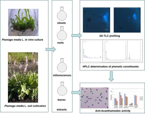
The protocol of efficient micropropagation of Plantago media L. was developed previously [19]. The shoots and roots of in vitro micropropagated plantlets were chosen for the experiments to compare whether raw materials produced by biotechnological methods are richer in selected secondary metabolites and have higher biological activity, in this case, acanthamoebic. Moreover, to compare the activity of extracts resulting from the presence and content of bioactive compounds, Plantago media was compared to the well-known and valued raw material of Plantago lanceolatae folium (Figure 1).
Chromatographic examination of extracts by 2D-TLC fingerprinting on cellulose plates (Table 1) exhibited a similar pattern of phenolic compounds in shoots and roots of the micropropagated plantlets. Phenylethanoid glycosides, such as acteoside and plantamajoside, were present, and no flavonoids could be detected. The phenylethanoids appeared under UV light as blue bands exhibiting strong blue fluorescence after spraying with NA. In turn, no yellow or brown bands changing color to brilliant yellow fluorescence under UV upon derivatization with aluminum chloride, characteristic for flavonoids, were observed. The flavonoids were apparent in the extracts of intact plants—P. media inflorescences and leaves and P. lanceolata leaves (Table 1).
The results of quantitative analyses performed using the HPLC-DAD method indicate differences in the content of individual compounds—phenylethanoids, flavonoids and phenolic acids—depending on the type of Plantago media L. raw material (Table 2).
The leaves of plants from the ground and from in vitro cultures possess the highest content of the sum of phenylethanoids as well as individual compounds—acteoside and isoacteoside. Moreover, the content of acteoside is more than two-fold higher in the leaves of the plant from the ground (6641.70 ± 12.06 mg/100 g DW) than from the shoot culture (3034.11 ± 15.54 mg/100 g DW). Roots from in vitro cultures contain lower contents of acteoside (1039.31 ± 9.18 mg/100 g) and isoacteoside (161.93 ± 6.42 mg/100 g), although more than inflorescences (655.13 ± 5.16 mg/100 g DW and 67.68 ± 2.50 mg/100 g DW, respectively), which are the least rich in compounds, both phenylethanoids and phenolic acids. In general, it can be observed that acteoside is the dominant compound in each of the raw materials of this species, regardless of the type of organ (leaf, root, or inflorescence) or origin (organs of natural plants or biomass from biotechnological production). Acteoside and isoacteoside (phenylethanoids) are present in raw materials in a much higher concentration than the sum of phenolic acids or the sum of flavonoids. The content of acteoside constitutes 87.76% and 84.15% (in vitro shoots and roots), 84.96% and 93.83% (in P. media inflorescences and leaves), and 93.60% (in P. lanceolata leaves) of the phenolics sum content. For comparison, the content of these compounds in Plantaginis lanceolatae folium, a well-known pharmacopoeial raw material, was also examined and it turned out that the leaves of P. media are richer in acteoside and isoacteoside than the leaves of P. lanceolata (Table 2).
The highest level of the sum of phenolic acids occurs, as with phenylethanoids, in the leaves, then the roots, and the least in the inflorescences of this species. In this case, the content of phenolic acids is higher in the leaves of the shoot culture (64.91 mg/100 g DW) than of the ground plant (53.42 mg/100 g DW). The predominant phenolic acid in the raw materials was protocatechuic acid (highest 24.76 ± 0.08 mg/100 g DW), gallic acid (highest 22.91 ± 0.08 mg/100 g DW), and vanillic acid (highest 15.86 ± 0.98 mg/100 g DW). The content of gallic acid and vanillic acid was two-fold higher in leaves from in vitro cultures than from a plant from the ground, while protocatechuic acid was two times more in leaves from shoot culture than in plants from the ground. The qualitative and quantitative composition in the range of phenolic acids of the leaves of P. media differed significantly from that in the leaves of P. lanceolata. In both raw materials, there was protocatechuic acid, but there was almost three times more in P. media than in P. lanceolata, in which ferulic and isoferulic acids were present (Table 2). In line with the 2D-TLC analysis, HPLC-DAD showed no flavonoids in the in-vitro-derived organs, in contrast to the soil-derived plants. These compounds were most abundant in P. media leaves (104.83 mg/100 g DW), followed by P. lanceolata leaves and P. media inflorescences (38.16 and 21.68 mg/100 g DW, respectively). P. media inflorescences and leaves contained the same compounds, the flavonols quercetin and isorhamnetin, and a flavone, apigenin, and the same principal constituent, isorhamnetin (15.02 ± 0.65 and 82.33 ± 4.82 mg/100 g DW, respectively). The P. lanceolata leaves shared quercetin and apigenin, but differed in the presence of the flavone luteolin and rutoside (quercetin diglycoside) as a major compound (23.48 ± 0.61 mg/100 g DW).
The results of the study indicated that the extracts obtained from Plantago, both from in vitro shoots and roots as well as from organs of intact plants, inhibited the growth of Acanthamoeba sp. trophozoites to varying degrees. The dependence of the effect on the extract concentration and treatment time was noted (Table 3 and Table 4) (Figure 2, Figure 3 and Figure 4).
The extract of leaves from shoot cultures was ineffective on Acanthamoeba trophozoites at the lowest concentration (1 mg/mL) because no statistical differences were observed compared to the control, regardless of the duration of treatment. Higher concentrations (5 mg/mL and 10 mg/mL) of the extract lowered the average number of trophozoites and, for them, on the fourth day of treatment, the growth inhibition was 63.89% and 81.44%, respectively (Table 3).
For the in vitro root extract, there is a clear tendency for the activity to be dependent on the time of exposure. On the second day, only the extract at the highest concentration worked; on the third day, extracts at the lowest and medium concentrations; and on the fourth day, extracts in all three concentrations were effective. However, the highest trophozoites growth inhibition was around 50–60%.
In the case of the inflorescences extract, the dependence of the effect on the extract concentration and treatment time was noted. The highest percentage of growth inhibition of Acanthamoeba trophozoites was noticed for the extract at a concentration of 10 mg/mL (61.47% on the second day, 69.75% on the third day, and 71.04% on the fourth day). The leaf extract showed antitrophozoic activity at all concentrations.
Similar to the root extract of P. media, there is a clear tendency for the activity of the leaf extract of P. lanceolata to be dependent on the time of exposure. The highest Acanthamoeba trophozoites growth inhibition was noticed after incubation with the extract at 10 mg/mL (on the second day 86.85%, on the third day 86.35%, and on the fourth day 86.27%) (Table 3).
The graphical comparison shown in Figure 3 suggested that IC50 values and the contents of phenolic constituents in the Plantago organs are inversely correlated. To check the nature of this correlation, the data were subjected to a Pearson test (Figure 4).
The analysis of the Pearson correlation coefficients (Figure 4) revealed the strongest correlation between IC50 and the content of the phenylethanoid glycosides sum and acteoside, followed by that of flavonoids sum, and the weakest correlation was found for the phenolic acids sum (mean r values were −0.8539 ± −0.84, −0.8506 ± −0.84, −0.7113 ± −0.68, and −0.6016 ± −0.58, respectively). The correlation with all phenolics sum was r −0.8541 ± −0.84, i.e., equal to that of phenylethanoid glycosides and very close to that of acteoside. The negative sign of the Pearson coefficients (Figure 4) proves that IC50 values and phenolics contents are inversely correlated—the higher the content, the stronger the activity.
4. Discussion
Acanthamoeba species have become recognized as dangerous agents leading to diseases including granulomatous amoebic encephalitis (GAE), amoebic keratitis (AK), and amoebic pneumitis (AP). The various synthetic anti-Acanthamoeba substances are often ineffective. Regarding the anti-parasitic activity, a plethora of raw materials and groups of bioactive compounds with amoebicidal or amoebistatic properties were studied including abietane diterpenoids from Salvia sclarea L. (with the highest activity of ferruginol—72%) [45]; essential oil from Pterocaulon polystachyum D.C. [46]; extracts from Artemisia argyi H.Lev&Vaniot [47] and Artemisia annua L. with the artemisinin as the most active compound [34]; Lonicera japonica Thunb. extract with its major constituent chlorogenic acid [48]; Buddleja cordata Tunht. extract rich in laminarin belonging to flavones [49]; a brew of green tea (Camelia sinensis (L.) Kuntze) rich in flavans [50]; extracts of Eryngium species [33,51]; Rubus chamaemorus L. extract with ellagic acid as the leading compound; Pueraria lobata (Willd.) Ohwi. extract with active isoflavones—puerarin and daidzein [32]; extracts of Teucrium species [52]; Centaurea species, Rhaponticum pulchrum Fisch. et Mey. and Tanacetum vulgare L. extracts with sesquiterpene lactones [53]; Lychnis flos-cuculi L. extracts with phytoecdysones [54]; and Chaenomeles japonica (Thunb.) Lindl. ex Spach extract with a high content of pentacyclic triterpenoids (mainly ursolic, oleanolic, and betulinic acids) [55].
Only amoebicidal substances are useful in the treatment of Acanthamoeba infections. Amoebostatic (change of trophozoites into cysts) substances are not suitable for use in therapy. The transformation of amoeba into cysts under the influence of the administered preparation is very unfavorable for the patient. The resulting cysts can survive in the patient’s tissues for a very long time and, under conditions favorable for the amoeba, they can cause the invasion to resume. Our task was to find a substance that would kill the trophozoites, but would not cause cyst formation. In the current study, no encystation (transformation of amoeba into cysts) was observed upon application of the tested extracts from Plantago.
In the present study, the highest anti-amoebic activity of the water fractions of Plantago media was recorded for concentrations of 5 and 10 mg/mL on the third and fourth day of treatment of trophozoites.
Leaf extracts from the crop with the highest content of acteoside had an anti-amoebic effect from the lowest concentration of 1 mg/mL and achieved the lowest IC50 value among the tested samples, while extracts from inflorescences and in vitro roots with the lowest content of acteoside also showed the highest IC50 index. Plantago lanceolata leaf extracts had a lower concentration of acteoside than P. media leaves and reached the lowest IC50 result, although only on the fourth day of the experiment, which, in turn, may be associated with a different profile of phenolic acids in these species. The presence of flavonoids in the composition does not seem to affect the strength of the antiprotozoal effect, which is confirmed by the particularly good result of this effect of in vitro shoot extracts devoid of these components.
The mechanism behind the antiprotozoal, exactly anti-leishmanial activity of acteoside has been found to be the inhibition of an arginase enzyme, leading to the inhibition of polyamine biosynthesis that is important to infectivity, and has been attributed to the catechol groups in this compound (two adjacent hydroxyl groups on each of the two phenyl rings) [28]. In our studies, the correlation between IC50 values and groups of phenolic secondary metabolites was highest for phenylethanoid glycosides and acteoside (R2 0.6248 and 0.6131, respectively). Although the inhibition of arginase has not been mentioned among modes of anti-acanthamoebic action [30], it is possible due to the ubiquitous occurrence of that enzyme [56]. The brews of green tea (Camelia sinensis), known for the high content of catechin derivatives possessing catechol and pyrogallol groups (e.g., EGCG—epigallocatechin 3-gallate), have exhibited pronounced anti-acanthamoebic activity [50]. The authors have hypothesized that the activity could be due to the inhibition of DHFR (dihydrofoliate reductase) and serine protease and metalloprotease enzymes resulting in the disruption of DNA synthesis and cytolysis of Acanthamoeba trophozoites, respectively [50].
When it comes to all the Plantago media extracts used, it can be seen that the lowest IC50, and thus the highest biological activity, have leaf extracts, both from ground plants (IC50 from 0.95 to 1.80 mg/mL) and obtained by biotechnological methods in vitro (3.30 to 5.50 mg/mL). A similar tendency was observed in the case of extracts from the leaves of shoot cultures of E. campestre and E. planum, for which IC50 was estimated to be lower than that for extracts from roots obtained in vitro [51]. As it turned out, this activity did not correlate with the content of polyphenols, which, for example, for Eryngium planum, was similar in roots and shoots obtained from micropropagated plants or with the content of triterpene saponins, which was impressively higher in the case of in-vitro-derived roots [55]. If we consider extracts from. C. japonica, IC50 was the lowest for extracts from leaves of shoots regenerated in vitro than from shoots of plants from natural sites, callus, or fruits [55]. The anti-acanthomoebic activity of the extracts clearly correlated with the content of pentacyclic triterpenoids [55]. Considering the anti-acanthoamoebic activity of L. flos-cuculi extracts, 80% methanol fractions from the herb had a higher IC50 value than those from roots or even biotechnologically obtained callus cells [54]. This relationship is probably not conditioned by the amount of main ecdysteroids, because, in the case of this species, the content of the sum of polypodine B and 20-hydroxyecdysone in the herb and roots is similar, while these compounds are not synthesized by callus cells [57]. Among aerial part extracts from plant species belonging to the Asteraceae family (C. bella, C. daghestanica, R. pulchrum, and T. vulgare), the lowest IC50 was calculated for C. bella, C. daghestanica, and T. vulgare, while IC50 for R. pulchrum was very high (>10 mg/mL). The amoebicidal effect of extracts may be attributed mainly to the sesquiterpene lactones, which are major compounds of four species from Asteraceae. The most common sesquiterpene lactones isolated from the investigated plants were guaianolides: cynaropicrin, janerin, and chlorojanerin [53]. On the other hand, the acanthamoebic activity of Origanum syriacum and O. laevigatum extracts seemed to be very low, even at a concentration of 32 mg/mL [58].
The above-discussed data on the anti-acanthamoebic activity of various plant species are compiled in the Table 5.
5. Conclusions
Our investigations show that Plantago media, either from in vitro cultures or soil cultivation, exerts an interesting effect against protozoan parasites such as Acanthamoeba sp. trophozoites in a manner dependent on the content of the main phenolic component, a phenylethanoid glycoside—acteoside (verbascoside). These findings imply that further investigations are needed to substantiate the role of acteoside, and the structurally related phenylethanoid glycosides, in the anti-acanthamoeba activity.
Author Contributions
Conceptualization, A.B. and M.K.; methodology, A.B., M.D., J.B. and A.S.; formal analysis, A.B., M.D., J.B. and A.S.; investigation, A.B., M.D., J.B. and A.S.; resources, A.B., M.D., J.B. and A.S.; data curation, A.B., M.D., J.B. and A.S.; writing—original draft preparation, M.K., A.B. and J.B.; writing—review and editing, M.K., A.B. and J.B.; visualization, M.K., A.B. and J.B.; supervision, M.K. and A.B. All authors have read and agreed to the published version of the manuscript.
Funding
This research received no external funding.
Institutional Review Board Statement
Not applicable.
Informed Consent Statement
Not applicable.
Data Availability Statement
Not applicable.
Conflicts of Interest
The authors declare no conflict of interest.
References
- POWO. Plants of the World Online. Facilitated by the Royal Botanic Gardens, Kew. 2023. Available online: https://powo.science.kew.org/taxon/urn:lsid:ipni.org:names:30001135-2 (accessed on 16 March 2023).
- POWO. Plants of the World Online. Facilitated by the Royal Botanic Gardens, Kew. 2023. Available online: https://powo.science.kew.org/taxon/urn:lsid:ipni.org:names:685411-1 (accessed on 10 March 2023).
- Fierascu, R.C.; Fierascu, I.; Ortan, A.; Paunescu, A. Plantago media L.—Explored and Potential Applications of an Underutilized Plant. Plants 2021, 10, 265. [Google Scholar] [CrossRef] [PubMed]
- Gonçalves, S.; Romano, A. The medicinal potential of plants from the genus Plantago (Plantaginaceae). Ind. Crops Prod. 2016, 83, 213–226. [Google Scholar] [CrossRef]
- European Pharmacopoeia. Ribwort leaf. In Plantaginis lanceolatae folium, 10th ed.; The Council of Europe: Strasbourg, France, 2019; p. 1599. [Google Scholar]
- Bogdanova, E.S.; Grebenkina, T.M.; Nesterov, V.N.; Rozentsvet, O.A. Biologically active compounds from representatives of the family Plantaginaceae. Chem. Nat. Compd. 2014, 50, 1001–1004. [Google Scholar] [CrossRef]
- Chiang, L.C.; Ng, L.T.; Chiang, W.; Chang, M.Y.; Lin, C.C. Immunomodulatory activities of flavonoids, monoterpenoids, triterpenoids, iridoid glycosides and phenolic compound of Plantago species. Planta Med. 2003, 69, 600–604. [Google Scholar] [CrossRef] [Green Version]
- Fleer, H.; Verspohl, E.J. Antispasmodic activity of an extract from Plantago lanceolata L. and some isolated compounds. Phytomedicine 2007, 14, 409–415. [Google Scholar] [CrossRef]
- Weryszko-Chmielewska, E.; Matysik-Woźniak, A.; Sulborska, A.; Rejdak, R. Commercially important properties of the genus Plantago. Acta Agron. 2012, 65, 11–20. [Google Scholar] [CrossRef] [Green Version]
- Rønsted, N.; Franzyk, H.; Molgaard, P.; Jaroszewski, J.W.; Jensen, S.R. Chemotaxonomy and evolution of Plantago L. Plant Syst. Evol. 2003, 242, 63–82. [Google Scholar] [CrossRef]
- Rønsted, N.; Göbel, E.; Franzyk, H.; Jensen, S.R.; Olsen, C.E. Chemotaxonomy of Plantago. Iridoid glucosides and caffeoyl phenylethanoid glycosides. Phytochemistry 2000, 55, 337–348. [Google Scholar] [CrossRef]
- Olennikov, D.N.; Tankhaeva, L.M.; Stolbikova, A.V.; Petrov, E.V. Phenylopropanoids and polysaccharides from Plantago depressa and Plantago media growing in Buryatia. Chem. Nat. Compd. 2011, 47, 165–169. [Google Scholar] [CrossRef]
- Andrzejewska-Golec, E. Taxonomic aspects of the iridoid glucosides occurring in the genus Plantago L. Acta Soc. Bot. Pol. 1997, 66, 201–205. [Google Scholar] [CrossRef] [Green Version]
- Taskova, R.; Evstatieva, L.; Handjieva, N.; Popov, S. Iridoid Patterns of Genus Plantago L. and Their Systematic Significance. Z. Nat. C 2002, 57, 42–50. [Google Scholar] [CrossRef] [PubMed] [Green Version]
- Jurišić Grubešić, R.; Srečnik, G.; Kremer, D.; Vuković Rodríguez, J.; Nikolić, T.; Vladimir-Knežević, S. Simultaneous RP–HPLC–DAD separation, and determination of flavonoids and phenolic acids in Plantago L. species. Chem. Biodiv. 2013, 10, 1305–1316. [Google Scholar] [CrossRef] [PubMed]
- Janković, T.; Zduni, G.; Beara, I.; Balog, K.; Pljevljakušić, D.; Stešsević, D.; Šavikin, K. Comparative study of some polyphenols in Plantago species. Biochem. Syst. Ecol. 2012, 42, 69–74. [Google Scholar] [CrossRef]
- Beara, I.N.; Lesjak, M.M.; Jovin, E.D.; Balog, K.J.; Anačkov, G.T.; Orčic, D.Z.; Mimica-Dukič, N.M. Plantain (Plantago L.) species as novel sources of flavonoid antioxidants. J. Agric. Food Chem. 2009, 57, 9268–9273. [Google Scholar] [CrossRef] [PubMed]
- Ji, H.; Hou, C.; Guo, X. Physicochemical properties, structures, bioactivities and future prospective for polysaccharides from Plantago L. (Plantaginaceae): A review. Int. J. Biol. Macromol. 2019, 135, 637–646. [Google Scholar] [CrossRef] [PubMed]
- Budzianowska, A.; Kikowska, M.; Małkiewicz, M.; Karolak, I.; Budzianowski, J. Phenylethanoid glycosides in Plantago media L. organs obtained in in vitro cultures. Acta Biol. Cracov. Ser. Bot. 2019, 61, 75–86. [Google Scholar] [CrossRef]
- Alipieva, K.; Korkina, L.; Orhan, I.E.; Georgiev, M.I. Verbascoside—A review of its occurrence, (bio)synthesis and pharmacological significance. Biotechnol. Adv. 2014, 32, 1065–1076. [Google Scholar] [CrossRef]
- Fu, G.; Pang, H.; Wong, Y.H. Naturally Occurring Phenylethanoid Glycosides: Potential Leads for New Therapeutics. Curr. Med. Chem. 2008, 15, 2592–2613. [Google Scholar] [CrossRef] [Green Version]
- Wu, L.; Georgiev, M.I.; Cao, H.; Nahar, L.; El-Seedi, H.R.; Sarker, S.D.; Xiao, J.; Lu, B. Therapeutic potential of phenylethanoid glycosides: A systematic review. Med. Res. Rev. 2020, 40, 2605–2649. [Google Scholar] [CrossRef]
- Xue, Z.; Yang, B. Phenylethanoid glycosides: Research advances in their phytochemistry, pharmacological activity and pharmacokinetics. Molecules 2016, 21, 991. [Google Scholar] [CrossRef]
- Bernardi, M.; Ghaani, M.R.; Bayazeid, O. Phenylethanoid glycosides as a possible COVID-19 protease inhibitor: A virtual screening approach. J. Mol. Model. 2001, 27, 341. [Google Scholar] [CrossRef]
- Verutani, S.; Beghelli, E.; Scalambra, E.; Malisardi, G.; Copetti, S.; Dal, T.R.; Baldisserotto, A.; Manfredini, S. Activity and stability studies of acteoside, a novel antioxidant, in dermo-cosmetic and pharmaceutical topical formulations. Molecules 2011, 16, 7068–7080. [Google Scholar] [CrossRef]
- Xiao, Y.; Ren, Q.; Wu, L. The pharmacokinetic property and pharmacological activity of acteoside: A review. Biomed Pharm. 2022, 153, 113296. [Google Scholar] [CrossRef] [PubMed]
- Khan, R.A.; Hossain, R.; Roy, P.; Jain, D.; Mohammad Saikat, A.S.; Roy Shuvo, A.P.; Akram, M.; Elbossaty, W.F.; Khan, I.N.; Painuli, S.; et al. Anticancer effects of acteoside: Mechanistic insights and therapeutic status. Eur. J. Pharmacol. 2022, 916, 174699. [Google Scholar] [CrossRef] [PubMed]
- Maquiaveli, C.C.; Lucon-Júnior, J.F.; Brogi, S.; Campiani, G.; Gemma, S.; Vieira, P.C.; Silva, E.R. Verbascoside inhibits promastigote growth and arginase activity of Leishmania amazonensis. J. Nat. Prod. 2016, 79, 1459–1463. [Google Scholar] [CrossRef] [PubMed]
- Maquiaveli, C.C.; Rochetti, A.L.; Fukumasu, H.; Vieira, P.C.; da Silva, E.R. Antileishmanial activity of verbascoside: Selective arginase inhibition of intracellular amastigotes of Leishmania (Leishmania) amazonensis with resistance induced by LPS plus IFN-γ. Biochem. Pharmacol. 2017, 127, 28–33. [Google Scholar] [CrossRef] [PubMed]
- Siddiqui, R.; Khan, N.A. Biology and pathogenesis of Acanthamoeba. Parasit Vectors 2012, 10, 5–6. [Google Scholar] [CrossRef] [Green Version]
- Derda, M.; Hadaś, E. The use of phytotherapy in diseases caused by parasitic protozoa. Acta Parasitol. 2015, 60, 1–8. [Google Scholar] [CrossRef]
- Derda, M.; Hadaś, E.; Thiem, B. Plant extracts as natural amoebicidal agents. Parasitol. Res. 2009, 104, 705–708. [Google Scholar] [CrossRef]
- Derda, M.; Thiem, B.; Budzianowski, J.; Wojt, W.; Wojtkowiak-Giera, A. The evaluation of the amebicidal activity of Eryngium planum extracts. Acta Pol. Pharm. 2013, 70, 1027–1034. [Google Scholar]
- Derda, M.; Hadaś, E.; Cholewiński, M.; Skrzypczak, Ł.; Grzondziel, A.; Wojtkowiak-Giera, A. Artemisia annua L. as a plant with potential use in the treatment of acanthamoebiasis. Parasitol. Res. 2016, 115, 1635–1639. [Google Scholar] [CrossRef] [PubMed] [Green Version]
- Elsheikha, H.M.; Siddiqui, R.; Khan, N.A. Drug discovery against Acanthamoeba infections: Present knowledge and unmeneeds. Pathogens 2020, 9, 405. [Google Scholar] [CrossRef] [PubMed]
- ESCOP Monographs. Plantaginis lanceolatae folium/herba. European Scientific Cooperation On Phytotherapy. In The Scientific Foundation for Herbal Medicinal Products, 2nd ed.; Thieme: Stuttgart, Germany, 2003; pp. 383–387. [Google Scholar]
- Harborne, J.B. Phytochemical Methods; Chapman and Hall: London, UK, 1998. [Google Scholar]
- Budzianowska, A.; Skrzypczak, L.; Budzianowski, J. Phenylethanoid glucosides from in vitro propagated plants and callus cultures of Plantago lanceolata. Planta Med. 2004, 70, 834–840. [Google Scholar] [CrossRef]
- Budzianowski, J.; Korzeniowska, K.; Chmara, E.; Mrozikiewicz, A. Microvascular protective activity of flavonoid glucuronides fraction from Tulipa gesneriana. Phytother Res. 1999, 13, 166–168. [Google Scholar] [CrossRef]
- Schlauer, J.; Budzianowski, J.; Kukulczanka, K.; Ratajczak, L. Acteoside and related phenylethanoid glycosides in Byblis liniflora Salisb. plants propagated in vitro and its systematic significance. Acta Soc. Bot. Pol. 2004, 73, 9–15. [Google Scholar] [CrossRef] [Green Version]
- Budzianowska, A.; Budzianowski, J. A new flavonoid, a new phenylethanoid glycoside and related compounds isolated from the inflorescences of Plantago lanceolata L. Nat. Prod. Res. 2022, 36, 3813–3824. [Google Scholar] [CrossRef]
- Markham, K.R. Techniques of Flavonoids Identification; Academic Press: London, UK, 1982. [Google Scholar]
- Szopa, A.; Kokotkiewicz, A.; Bednarz, M.; Luczkiewicz, M.; Ekiert, H. Studies on the accumulation of phenolic acids and flavonoids in different in vitro culture systems of Schisandra chinensis (Turcz.) Baill. using a DAD-HPLC method. Phytochem. Lett. 2017, 20, 462–469. [Google Scholar] [CrossRef]
- Szopa, A.; Kokotkiewicz, A.; Kubica, P.; Banaszczak, P.; Wojnatowska-Krośniak, A.; Krośniak, M.; Marzec-Wróblewska, U.; Badura, A.; Zagrodzki, P.; Buciński, A.; et al. Comparative analysis of different groups of phenolic compounds in fruit and leaf extracts of Aronia sp.: A. melanocarpa, A. arbutifolia, and A.×prunifolia and their antioxidant activities. Eur. Food Res. Technol. 2017, 243, 1645–1657. [Google Scholar] [CrossRef] [Green Version]
- Kuźma, Ł.; Derda, M.; Hadaś, E.; Wysokińska, H. Abietane diterpenoids from Salvia sclarea transformed roots as growth inhibitors of pathogenic Acanthamoeba spp. Parasitol. Res. 2015, 114, 323–327. [Google Scholar] [CrossRef]
- Sauter, I.P.; dos Santos, J.C.; Apel, M.A.; Cibulski, S.P.; Roehe, P.M.; von Poser, G.L.; Rott, M.B. Amoebicidal activity and chemical composition of Pteroculon polystachyum (Asteraceae) essential oil. Parasitol. Res. 2011, 109, 1367–1371. [Google Scholar] [CrossRef]
- Kolören, O.; Kolören, Z.; Şekeroğlu, Z.A.; Colayvaz, M.; Karanis, P. Amoebicidal and amoebistatic effects of Artemisia argyi methanolic extracts on Acanthamoeba castellanii trophozoites and cysts. Acta Parasitol. 2019, 64, 63–70. [Google Scholar] [CrossRef] [PubMed]
- Mahboob, T.; Azlan, A.-M.; Tan, T.-C.; Samudi, C.; Sekaran, S.D.; Nissapatorn, V.; Wiart, C. Anti-encystment and amoebicidal activity of Lonicera japonica Thunb. and its major constituent chlorogenic acid in vitro. Asian Pac. J. Trop. Med. 2016, 9, 866–871. [Google Scholar] [CrossRef] [PubMed] [Green Version]
- Rodríguez-Zaragoza, S.; Ordaz, C.; Avila, G.; Muñoz, J.L.; Arciniegas, A.; Romo de Vivar, A. In vitro evaluation of the ame-bicidal activity of Buddleia cordata (Loganiaceae, H.B.K.) on several strains of Acanthamoeba. J. Ethnopharmacol. 1999, 66, 327–334. [Google Scholar] [CrossRef]
- Fakae, L.B.; Stevenson, C.W.; Zhu, X.-Q.; Elsheikha, H.M. In vitro activity of Camellia sinensis (green tea) against trophozoites and cysts of Acanthamoeba castellanii. Int. J. Parasitol. Drugs Drug Resist. 2020, 13, 59–72. [Google Scholar] [CrossRef] [PubMed]
- Kikowska, M.; Chanaj-Kaczmarek, J.; Derda, M.; Budzianowska, A.; Thiem, B.; Ekiert, H.; Szopa, A. The evaluation of phenolic acids and flavonoids content and antiprotozoal activity of Eryngium species biomass produced by biotechnological methods. Molecules 2020, 27, 363. [Google Scholar] [CrossRef]
- Tepe, B.; Malatyali, E.; Degerli, S.; Berk, S. In vitro amoebicidal activities of Teucrium polium and T. chamaedrys on Acanthamoeba castellanii trophozoites and cyst. Parasitol. Res. 2011, 110, 1773–1778. [Google Scholar] [CrossRef]
- Hadaś, E.; Derda, M.; Nawrot, J.; Nowak, G.; Thiem, B. Evaluation of the Amoebicidal Activities of Centaurea bella, Centaurea daghestanica, Rhaponticum pulchrum and Tanacetum vulgare against Pathogenic Acanthamoeba spp. Acta Pol. Pharm. 2017, 74, 1827–1832. Available online: https://www.ptfarm.pl/download/?file=File%2FActa_Poloniae%2F2017%2F6%2F1827.pdf (accessed on 10 April 2023).
- Maliński, M.P.; Budzianowski, J.; Kikowska, M.; Derda, M.; Jaworska, M.M.; Młynarczyk, D.T.; Szukalska, M.; Florek, E.; Thiem, B. Two ecdysteroids isolated from micropropagated Lychnis flos-cuculi and the biological activity of plant material. Molecules 2021, 26, 904. [Google Scholar] [CrossRef]
- Kikowska, M.; Derda, M.; Thiem, B.; Włodarczyk, A.; Długaszewska, J.; Stochmal, A.; Żuchowski, J.; Hadaś, E. Evaluation of antiamoebic and antimicrobial activities in vitro of Chaenomeles japonica (Thunb.) Lindl. ex spach extracts. Acta Biol. Cracov. Ser. Bot. 2019, 61, 47–58. [Google Scholar] [CrossRef]
- Caldwell, R.B.; Toque, H.A.; Narayanan, S.P.; Caldwell, R.W. Arginase: An old enzyme with new tricks. Trends Pharmacol. Sci. 2015, 36, 395–405. [Google Scholar] [CrossRef] [Green Version]
- Maliński, M.P.; Kikowska, M.; Kruszka, D.; Napierała, M.; Florek, E.; Sliwinska, E.; Thiem, B. Various in vitro systems of Ragged Robin (Lychnis flos-cuculi L.): A new potential source of phytoecdysteroids? Plant Cell Tiss. Organ Cult. 2019, 139, 39–52. [Google Scholar] [CrossRef] [Green Version]
- Degerli, S.; Tepe, B.; Celiksoz, A.; Berk, S.; Malatyali, E. In vitro amoebicidal activity of Origanum syriacum and Origanum laevigatum on Acanthamoeba castellanii cysts and trophozoites. Exp. Parasitol. 2012, 131, 20–24. [Google Scholar] [CrossRef] [PubMed]
Figure 1.
Plant materials used for the investigations: Plantago media in soil cultivation (a) with zoomed inflorescences (d) and leaves (e); and P. media in-vitro-derived leaves (b) and roots (c).
Figure 1.
Plant materials used for the investigations: Plantago media in soil cultivation (a) with zoomed inflorescences (d) and leaves (e); and P. media in-vitro-derived leaves (b) and roots (c).

Figure 2.
Effect of the extracts from Plantago media in vitro shoots (a), in vitro roots (b), inflorescences (c) and leaves (d), and Plantago lanceolata leaves (e) on the growth of Acanthamoeba sp.
Figure 2.
Effect of the extracts from Plantago media in vitro shoots (a), in vitro roots (b), inflorescences (c) and leaves (d), and Plantago lanceolata leaves (e) on the growth of Acanthamoeba sp.

Figure 3.
Comparison of IC50 values of anti-acanthamoebic activity (a) with the content of phenolic secondary metabolites in the investigated organs of Plantago (b).
Figure 3.
Comparison of IC50 values of anti-acanthamoebic activity (a) with the content of phenolic secondary metabolites in the investigated organs of Plantago (b).

Figure 4.
Correlations between IC50 values and the content of phenylethanoid glycosides (a), acteoside (b), phenolic acids (c), flavonoids in all materials (d), flavonoids without consideration of in vitro-derived materials (e), and phenolics sum (phenylethanoid glycosides + phenolic acids + flavonoids) (f). * The Pearson correlation coefficients are statistically significant at two-tailed p < 0.05.


Table 1.
TLC-fingerprinting analysis of phenolic compounds in Plantago media.
| Material (Water Fraction of an Extract) | 2D-TLC Cellulose (Adsorbent: Cellulose; Mobile Phases: I: BAW 4:1:5, II: 15%AcOH; Detection—UV 365 nm) | Flavonoids | Phenylethanoid Glycosides |
|---|---|---|---|
| Plantago media in vitro shoots |  | Absent | Present |
| Plantago media in vitro roots |  | Absent | Present |
| Plantago media inflorescences |  | Present | Present |
| Plantago media leaves |  | Present | Present |
| Plantago lanceolata leaves |  | Present | Present |
Fla—flavonoids (brown spots); and PhG—phenylethanoid glycosides (blue spots).
Table 2.
The content of phenolic compounds [mg/100 g DW ± SD] in extracts of biomass from in vitro cultures (shoots and roots) and organs of plants from the field cultivation.
Table 2.
The content of phenolic compounds [mg/100 g DW ± SD] in extracts of biomass from in vitro cultures (shoots and roots) and organs of plants from the field cultivation.
| Compounds | P. media In Vitro Shoots | P. media In Vitro Roots | P. media Inflorescences | P. media Leaves | P. lanceolata Leaves |
|---|---|---|---|---|---|
| Phenolic acids | |||||
| Gallic acid | 22.91 ± 0.08 a | 17.14 ± 0.09 b | 10.97 ± 0.12 d | 11.40 ± 0.13 c | - |
| Protocatechuic acid | 12.24 ± 0.13 b | 1.08 ± 0.01 d | 7.72 ± 0.15 c | 24.76 ± 0.08 a | 8.99 ± 1.26 c |
| Vanillic acid | 15.86 ± 0.98 a | 5.26 ± 0.21 c | 4.53 ± 0.67 d | 8.00 ± 0.50 b | - |
| Caffeic acid | 5.96 ± 1.41 a | 3.51 ± 0.98 bc | 1.28 ± 0.04 d | 2.49 ± 0.68 c | - |
| Syringic acid | 7.94 ± 1.55 a | 6.87 ± 0.99 a | 2.12 ± 0.09 b | 6.77 ± 0.34 a | - |
| Ferulic acid | - | - | - | - | 7.28 ± 1.34 |
| Isoferulic acid | - | - | - | - | 34.86 ± 2.13 |
| Sum | 64.91 | 33.86 | 26.62 | 53.42 | 51.13 |
| Phenylethanoids | |||||
| Acteoside | 3034.11 ± 15.54 c | 1039.31 ± 9.18 d | 655.13 ± 5.16 e | 6641.70 ± 12.06 a | 4015.60 ± 40.2 b |
| Isoacteoside | 358.15 ± 7.03 a | 161.93 ± 6.42 d | 67.68 ± 2.50 e | 278.55 ± 4.64 b | 185.06 ± 8.08 c |
| Sum | 3392.26 | 1201.24 | 722.81 | 6920.25 | 4200.66 |
| Flavonoids | |||||
| Quercetin | - | - | 2.00 ± 0.12 | 11.95 ± 0.58 | 2.24 ± 0.68 |
| Isorhamnetin | - | - | 15.02 ± 0.65 | 82.33 ± 4.82 | - |
| Apigenin | - | - | 4.66 ± 0.29 | 10.57 ± 0.43 | 11.18 ± 0.39 |
| Luteolin | - | - | - | - | 1.23 ± 0.61 |
| Rutoside | - | - | - | - | 23.48 ± 0.61 |
| Sum | 0 | 0 | 21.68 | 104.85 | 38.16 |
| Sum of all compounds | 3457.17 | 1235.10 | 771.11 | 7078.52 | 4289.95 |
Mean values within a row with the same letter are not significantly different at p < 0.05 using Duncan’s multiple range test—the first letter of the alphabet for the highest values, the next for statistically significant decreasing values.
Table 3.
Effect of extracts from Plantago media biomass from in vitro culture (shoots and roots) and organs of plants from the cultivation on inhibition of Acanthamoeba trophozoites during four-day treatment.
Table 3.
Effect of extracts from Plantago media biomass from in vitro culture (shoots and roots) and organs of plants from the cultivation on inhibition of Acanthamoeba trophozoites during four-day treatment.
| Plant Material | Extract Concentration | Duration of Treatment [Days] | |||||
|---|---|---|---|---|---|---|---|
| 2nd Day | 3rd Day | 4th Day | |||||
| MN ± SD | % GI | MN ± SD | % GI | MN ± SD | % GI | ||
| Plantago media | control | 12.44 ± 2.22 | 0 | 22.94 ± 3.39 | 0 | 28.38 ± 4.33 | 0 |
| in vitro shoots | 1 mg/mL | 12.18 ± 2.77 | 2.10 | 18.41 ± 4.31 | 19.75 | 21.82 ± 6.80 | 23.11 |
| 5 mg/mL | 6.33 ± 1.98 * | 49.06 | 11.42 ± 5.07 * | 50.22 | 10.25 ± 3.47 * | 63.89 | |
| 10 mg/mL | 2.94 ± 1.61 * | 76.27 | 4.20 ± 2.14 * | 81.70 | 5.27 ± 5.66 * | 81.44 | |
| Plantago media | control | 12.44 ± 2.22 | 0 | 18.94 ± 3.39 | 0 | 27.38 ± 4.33 | 0 |
| in vitro roots | 1 mg/mL | 9.18 ± 2.55 | 26.21 | 13.00 ± 2.94 | 31.37 | 16.67± 1.25 * | 39.12 |
| 5 mg/mL | 8.83 ± 1.67 | 29.02 | 10.25 ± 3.49 * | 45.89 | 13.91 ± 4.46 * | 49.20 | |
| 10 mg/mL | 6.03 ± 1.91 * | 51.45 | 9.09 ± 2.35 * | 52.01 | 12.00 ± 2.52 * | 56.18 | |
| Plantago media | control | 12.12 ± 2.82 | 0 | 26.44 ± 6.04 | 0 | 28.00 ± 5.35 | 0 |
| inflorescences | 1 mg/mL | 10.76 ± 1.92 | 11.23 | 25.00 ± 4.97 | 5.45 | 26.27 ± 3.22 | 6.18 |
| 5 mg/mL | 9.83 ± 2.65 | 18.90 | 16.33 ± 3.73 * | 38.24 | 16.42 ± 4.35 * | 41.36 | |
| 10 mg/mL | 4.67 ± 2.38 * | 61.47 | 8.00 ± 2.47 * | 69.75 | 8.10 ± 3.97 * | 71.04 | |
| Plantago media | control | 12.12 ± 2.82 | 0 | 23.44 ± 6.04 | 0 | 32.00 ± 5.35 | 0 |
| leaves | 1 mg/mL | 6.00 ± 1.97 * | 50.50 | 12.33 ± 3.89 * | 47.40 | 17.67 ± 5.03 * | 44.79 |
| 5 mg/mL | 5.91 ± 1.73 * | 51.24 | 10.25 ± 3.92 * | 56.28 | 14.50 ± 4.03 * | 54.69 | |
| 10 mg/mL | 5.05 ± 2.39 * | 58.34 | 8.81 ± 3.43 * | 62.42 | 12.40 ± 3.88 * | 61.25 | |
| Plantago lanceolata | control | 10.11 ± 2.05 | 0 | 25.72 ± 4.36 | 0 | 38.39 ± 7.77 | 0 |
| leaves | 1 mg/mL | 9.76 ± 2.60 | 3.47 | 18.67 ± 4.88 | 27.42 | 14.00 ± 5.18 * | 63.54 |
| 5 mg/mL | 8.67 ± 1.97 | 14.25 | 16.35 ± 4.83 * | 26.57 | 10.25 ± 3.47 * | 74.31 | |
| 10 mg/mL | 1.33 ± 0.94 * | 86.85 | 3.51 ± 2.32 * | 86.35 | 5.27 ± 5.66 * | 86.27 | |
* p < 0.05, statistically significant difference in comparison with the control during the same time interval; n = 18. MN—mean number of trophozoites, and GI—growth inhibition.
Table 4.
Determination of IC50 [mg/mL] for the studied extracts of P. media and P. lanceolata.
| IC50 | P. media In-Vitro -Derived Shoots | P. media In-Vitro -Derived Roots | P. media Inflorescences | P. media Leaves | P. lanceolata Leaves |
|---|---|---|---|---|---|
| 2nd day | 5.20 | 9.70 | 8.70 | 0.95 | 7.65 |
| 3rd day | 5.00 | 7.60 | 6.80 | 1.25 | 7.25 |
| 4th day | 3.30 | 5.30 | 6.30 | 1.80 | 0.55 |
IC50—the half-maximum anti-amoebic inhibitory concentration.
Table 5.
IC50 value for the studied discussed plant species.
| Plant Species | IC50 on 2nd Day | References |
|---|---|---|
| Plantago media L. | Water-soluble fr of MeOH extract from leaves of intact plants, 0.95 mg/mL Water-soluble fr of MeOH extract leaves of in vitro plants, 5.20 mg/mL | This work |
| Salvia sclarea L. | Ferruginol 0.05 mg/mL Salvipisone 0.4 mg/mL | [45] |
| Pterocaulon polystachyum DC | Essential oil 10 mg/mL | [46] |
| Artemisia argyi Levl. et Vant. | MeOH extract 37.4 mg/mL (8 h) | [47] |
| Artemisia annua L. | MeOH extract 12.5 mg/mL Artemisinin 0.12 mg/mL | [34] |
| Buddleya cordata H.B.K. | N.R. amoebostatic at 32 mg/mL | [49] |
| Camelia sinensis (L.) Kuntze | N.R. 50% brew | [50] |
| Eryngium planum L. | N.R. 50% inh. 5 mg/mL | [33,51] |
| Rubus chamaemorus L. | MeOH extract > 0.05 mg/mL | [32] |
| Pueraria lobata (Willd.) | MeOH extract > 0.01 mg/mL | [32] |
| Solidago virgaurea L. | MeOH extract > 0.01 mg/mL | [32] |
| Solidago graminifolia Elliott. | MeOH extract > 0.05 mg/mL | [32] |
| Teucrium polium L. | MeOH extract 60% inh. 16 mg/mL | [52] |
| Teucrium chamaedrys L. | MeOH extract 53% inh. 8 mg/mL | [52] |
| Lychnis flos-cuculi L. | 40% fr MeOH herb extract > 1.0 mg/mL 80% fr MeOH herb extract > 1.0 mg/mL 40% fr MeOH root extract > 5.0 mg/mL 80% fr MeOH root extract > 0.5 mg/mL 20-hydroxyecdysone > 0.5 mg/mL Polypodine B > 0.5 mg/mL | [54] |
| Chaenomeles japonica L. | EtOH fruit extract 6.25 mg/mL EtOH leaves extract 1.80 mg/mL EtOH leaves (in vitro) extract 0.3 mg/mL EtOH callus extract 2.30 mg/mL | [55] |
| Centaurea bella Trautv. | EtOH extract 0.61 mg/mL | [53] |
| Centaurea daghestanica Sosn. | EtOH extract 0.86 mg/mL | [53] |
| Rhaponticum pulchrum Fisch. Et Mey | EtOH extract > 10 mg/mL | [53] |
| Tanacetum vulgare L. | EtOH extract 0.64 mg/mL | [53] |
IC50—the half-maximum anti-amoebic inhibitory concentration; N.R.—not reported; and Inh.—inhibition.
Disclaimer/Publisher’s Note: The statements, opinions and data contained in all publications are solely those of the individual author(s) and contributor(s) and not of MDPI and/or the editor(s). MDPI and/or the editor(s) disclaim responsibility for any injury to people or property resulting from any ideas, methods, instructions or products referred to in the content. |
© 2023 by the authors. Licensee MDPI, Basel, Switzerland. This article is an open access article distributed under the terms and conditions of the Creative Commons Attribution (CC BY) license (https://creativecommons.org/licenses/by/4.0/).
Share and Cite
MDPI and ACS Style
Budzianowska, A.; Derda, M.; Budzianowski, J.; Szopa, A.; Kikowska, M. Comparative Study of Plantago media Extracts in the Treatment of Acanthamoeba sp. Trophozoites. Appl. Sci. 2023, 13, 7075. https://doi.org/10.3390/app13127075
AMA Style
Budzianowska A, Derda M, Budzianowski J, Szopa A, Kikowska M. Comparative Study of Plantago media Extracts in the Treatment of Acanthamoeba sp. Trophozoites. Applied Sciences. 2023; 13(12):7075. https://doi.org/10.3390/app13127075
Chicago/Turabian StyleBudzianowska, Anna, Monika Derda, Jaromir Budzianowski, Agnieszka Szopa, and Małgorzata Kikowska. 2023. "Comparative Study of Plantago media Extracts in the Treatment of Acanthamoeba sp. Trophozoites" Applied Sciences 13, no. 12: 7075. https://doi.org/10.3390/app13127075
Note that from the first issue of 2016, this journal uses article numbers instead of page numbers. See further details here.