Article

Isolation, Identification and Pollution Prevention of Bacteria and Fungi during the Tissue Culture of Dwarf Hygro (Hygrophila polysperma) Explants

Marine Science and Engineering College, Qingdao Agricultural University, Qingdao 266237, China
*
Author to whom correspondence should be addressed.
Microorganisms 2022, 10(12), 2476; https://doi.org/10.3390/microorganisms10122476
Submission received: 1 November 2022 / Revised: 8 December 2022 / Accepted: 12 December 2022 / Published: 15 December 2022

Abstract

:
Microbial contamination causes serious damage in plant tissue culture, and attention is always being paid regarding how to control and prevent the unwanted pollution. Dwarf hygro (Hygrophila polysperma) is a popular ornamental aquatic plant and its tissue culture has been reported, but the microbial pollution and the cure of microbial pollution was unknown. In this study, a number of bacteria and fungi were isolated from contaminants in MS culture media. Based on the 16S rDNA and ITS sequencing, it was identified that fifteen bacteria belong to Bacillus, Enterobacter, Pantoea, Kosakonia, Ensifer and Klebsiella, and three fungi belong to Plectosphaerella, Cladosporium and Peniophora, respectively. In addition, some drugs were further tested to be free of the bacteria and fungi pollution. The results revealed that 10 μg/mL of kanamycin, 5 μg/mL of chloramphenicol, and 0.015625% potassium sorbate could be applied jointly in MS media to prevent the microbial pollution, and the survival rate of H. polysperma explants was highly improved. This study reveals the bacteria and fungi species from the culture pollution of H. polysperma and provides a practical reference for optimizing the tissue culture media for other aquatic plants.

Graphical Abstract

1. Introduction

Plenty of aquatic species are naturally propagated at a long cultivation period, or subjected to unstable growth because of water transmitted diseases and variable environmental conditions [1]. However, planting the tissue culture of aquatic plants could break these limitations. Under artificial control, the explants from leaves and shoots, embryos or protoplasts were cultured on sterile media in vitro, and further, newly differentiated into a whole plant. In tissue culture, explants could be reproduced with high speed and heredity stability [2,3,4]. The success of in vitro plant culture was contributed to many biotic or abiotic factors, including the explants’ physiological status, the favorable culture media and ecological factors, etc. [5]. Among them, the control of microbial contamination was crucial. When the frequent pollution occurred in culture media, the explants could become withered and even die, and finally, it failed to be in progress of the tissue culture. Multiple factors caused the microbial pollution of plant tissue culture. In vitro, the incomplete sterilization, culture rooms and transfer fields are all possible contaminated sources [6]. In vivo, plants naturally keep a symbiotic relationship with diverse microorganisms, described as endophytes, including bacteria, fungi, and archaea [7,8,9]. Increasingly, a variety of endophytes were reported on high land plants, including Bacillus, Pseudomonas, Enterobacter, Rhizobium and Klebsiella, Agrobacterium, Methylobacterium spp., etc. [10,11,12,13,14]. Few were reported in aquatic plants, and it was yet unknown about the endophytes. Early in 1976, some mesophilic bacteria were identified from the purchased ornamental aquarium plants from retail outlets, and the dominant bacteria was Pseudomonas aeruginosa (P. aeruginosa) [15]. Recently, various drugs had been tested to avoid the pollution, including antibiotics, chemical compounds, Chinese traditional medicines, and nano materials [3,5,6,16]. However, these studies were mainly focused on high terrestrial plants [17,18,19]. There are few reports on the pollution and prevention of tissue culture in aquatic plants.
Aquatic plants are broadly defined as plants that undergo chloroxygenic photosynthesis, containing numerous single-cell and multicellular species with abundant populations [20]. They play ecological and functional roles in various aspects, such as assimilating excessive nutrients (e.g., nitrogen and phosphorus) in eutrophic waters [21,22], removing heavy metals and nanoparticles in wastewater [23,24,25,26,27], and maintaining mutual benefit with aquatic animals [28], as well as future biofuels [29] and medicinal plants [30,31,32,33]. Importantly, most of them were beautiful ornamental plants in the aquarium, with high commercial value [2,3,4,34,35]. Hygrophila polysperma (or Hygrophila rubella), known as dwarf hygro, belongs to Acanthaceae, the Hygrophila genus. H. polysperma has lanceolar leaves, sawtooth edge and clear veins, and leaves of plants often maintain a variable color; red at the top and green at the middle and base, depending on high light intensity and abundant nutrients. As an emergent macrophyte, it was usually chosen as a middle or background grass in aquariums. It is not only a popular aquarium plant, but also a medicinal plant and bioindicator for the control of algae and toxicities, it being in increasing market demand [3,31]. The tissue culture technique is an indispensible approach for the rapid propagation of H. polysperma. However, the microbial contamination frequently emerged in tissue culture of H. polysperma, and caused high mortality of explants. As previously reported, adventitious shoots of H. polysperma were regenerated in Murashige and Skoog (MS) culture media, and Amoklavin was applied for preventing the bacterial pollution [3]. Yet, it could not inhibit the fungi growth and was not convenient to acquire from the local farm in China. Thus, this study is aimed to reveal the bacteria and fungi species from the contaminants in culture media and further to seek the drugs to control them effectively for the success of tissue culture in H. polysperma.

2. Materials and Methods

2.1. Plant Material and Culture Conditions

H. polysperma were purchased from a local aquatic plant farm in Shandong province, China. Five-six cm long twigs of H. polysperma containing 5–6 nodes from plants with attached leaves were firstly washed for 5 min under tap water. In the clean bench, the leaves were removed from the twigs and the twigs were cut into the 4–5 cm long segments, containing 2–3 nodes, then it was surface-sterilized with 75% alcohol for 30 s, and rapidly transferred into 2% NaClO solution for 10 min, followed by rinsing three times with sterilized distilled water via continuous stirring. Subsequently, the sterilized twigs were cut into 0.5–1 cm explants, and cultured in 240 mL culture vessels (6 × 9 cm) containing MS medium for several weeks [36], which was supplemented with 3% sucrose, 1.0 mg/L of 6-KT and 1.0 mg/L of IBA, and solidified with 0.65% agar with the pH adjusted to 5.8 ± 0.1 before autoclaving (118 kPa atmospheric pressure, 121 °C for 20 min). Each culture vessel contained two-four shoot explants. All cultures were incubated at 26 ± 1 °C and 60–70% humidity under a 16 h light photoperiod (5000 lux) using white fluorescent lights in the climatic chamber (Ningbo, China).

2.2. Isolation and Identification of Bacteria

Based on the phenotypical observation, the bacterial contaminants, frequently appeared in MS media within a month. To culture the appearing bacteria as much as possible, bacteria were streaked with inoculation loops on the common microbial media, Luria-Bertani (LB) and tryticase soy broth (TSB) agar plates to incubate at 26 °C for 48 h, respectively. Then, single bacterial colonies were selected and restreaked on their preferential media again. When the morphology of all colonies on agar plates was identical, and the pure culture of bacterial isolate was obtained, all isolates were maintained separately in LB or TSB broth with 15% glycerol at −80 °C.

2.3. Isolation and Identification of Fungi

Several fungi also appeared in MS media. Based on the phenotypical observation, the typical fungi were chosen and inoculated on potato dextrose agar (PDA) plates, and then incubated at 26 °C for 72 h. After several inoculations, the fungal pure culture was obtained and stored on PDA plates at 4 °C until use.
The fungal shapes were detected by the scotch cellophane tape method using a lacto-phenol cotton blue (LPCB) solution to stain the fungi blue. One cellophane tape containing fungal sample was pressed against the surface of the glass slide, while another cellophane tape was pasted at its non-ground glass end. Then a drop of LPCB was placed on the side where the tape crosses. After several minutes, LPCB was permeated into the fungal bodies, and the fungal morphology was observed with a microscope and pictures were taken.

2.4. Molecular Analysis of Bacteria and Phylogenetic Tree Construction

The bacterial cells were washed twice with sterile phosphate buffer (pH 7.4), and genomic DNA were extracted using a FastPure® bacteria DNA isolation mini kit (Vazyme, Nanjing, China) according to the manufacturer’s protocol and stored at −20 °C until use, which was used as a template for PCR reaction. The PCR reaction contained 10.0 μL of 2 × Taq PCR Mastermix, 0.4 μL each of 10 μM sense primer (27F) and anti-sense primer (1492R), 1.0 μL of 500 ng of template DNA, in a final volume made up to 20 μL with sterile distilled water. PCR was performed for all isolates to amplify the 16S rDNA gene using the universal primers 27F (Sense primer, 5′-AGAGTTGATCATGGCTCA-3′) and 1492R (Anti-sense primer, 5′-GGTTCACTTGTTACGACTT-3′), as described previously [37]. The amplifications were carried out in a thermal cycler (BioRad, USA), with a pre-cycle denaturation at 95 °C for 5 min followed by 34 serial cycles of 94 °C for 30 s, 55 °C for 30 s, and 72 °C for 1 min, with a final extension at 72 °C for 7 min. The PCR products were analyzed by 1.2% (w/v) agarose gel (containing GelRed) electrophoresis in 1% tris–acetic acid–EDTA buffer. Gels were visualized and photographed under UV illumination. The PCR products were sent to the Ruibo Xingke biotechnology Co., Ltd. (Qingdao, China) and conducted by Sanger sequencing. Then, the 16S rDNA sequences were submitted to the NCBI GenBank database and blasted using the BLASTn algorithm. The identification of a specie of bacteria was more than 97% nucleotide identity for the 16S rDNA sequence.
A phylogenetic tree was created using the multi-sequence alignment generated via the CLUSTAL X program in Mega 6.0 by the neighbour-joining method [38]. The genetic distances matrix was obtained using Kimura’s two-parameter model [39], and bootstrap values from 1000 replicates are displayed as percentages.

2.5. Molecular Analysis of Fungi

The fungi hyphae were collected and grounded with sterile glass bead, and genomic DNA was extracted using a fungal DNA isolation Kit (Solarbio, Beijing, China). DNA as template was used for the PCR reaction to amplify the internal transcribed spacer (ITS) sequence of fungal ribosomal genes [40], using the primers of ITS-F (Sense primer, 5′-TCCGTAGGTGAACCTGCGG-3′) and ITS-R (Anti-sense primer, 5′-TCCTCCGCTTATTGATATGC-3′), followed by the similar protocol described as above in Section 2.4.

2.6. Screening Test for Bacterial Inhibition

Bacteria were grown at 26 °C in LB broth for 24 h. Bacterial growth was assessed for two weeks on LB broth and agar supplemented with 50 μg/mL of kanamycin (Kan), 100 μg/mL of ampicillin (Amp), 250 μg/mL of erythromycin (Ery), 200 μg/mL of spectinomycin (Spc), or 25 μg/mL of chloramphenicol (Chl) at 26 °C, respectively. The antibiotic tolerance levels were examined and recorded as sensitive (−) or resistant (+) accordingly.

2.7. Screening Test for Fungal Inhibition

The fungi growth was assessed for two weeks on PDA agar plates supplemented with 0.015625% (w/v), 0.03125% (w/v), 0.0625% (w/v), 0.1% (w/v), 0.125% (w/v), 0.25% (w/v), 0.5% (w/v) and 1% (w/v) potassium sorbate (PS) at 26 °C, respectively. Equally, a series concentration of sodium benzoate (SB) or sodium diacetate (SD) solution, as well as nanoscale silver (nano-Ag) was tested for the inhibition of fungi, respectively.

2.8. The Influences of the Antimicrobial Agents on Explants

The effects of antimicrobial agents on explants were tested. On the one hand, 50 μg/mL of Kan and 25 μg/mL of Chl (Kan + Chl), 1/2 (Kan + Chl), 1/5 (Kan + Chl) and 1/10 (Kan + Chl) were added into MS media, then the inhibition of bacteria was examined and the explants’ growth was observed for two weeks. On the other hand, a series of concentration (0.015625%, 0.03125%, 0.0625%, 0.1%, 0.2% and 0.5%) of PS, SB and SD were investigated on the inhibition of fungi and influences on the explants’ growth for one month. Subsequently, the appropriate concentration of antimicrobial agents was determined. Then, the explants or seedlings were grown in MS media supplemented with the above appropriate concentration of drugs. Finally, the seedling was transplanted into the soil of the aquarium, and its growth was recorded and photos were taken for two months.

3. Results

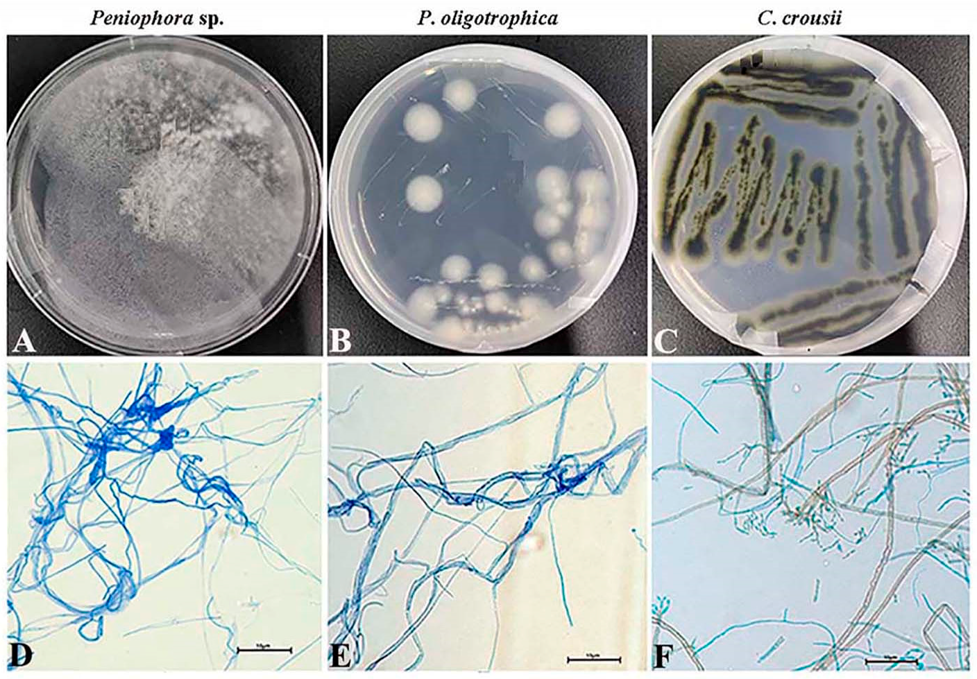
3.1. Phenotypic Profiles of the Microbial Contaminants

Currently, via the tissue culture technique, H. polysperma plantlets could be rapidly propagated using the young leaf and stem explants in agar-gelled MS media. Although H. polysperma explants were sterilized with 75% alcohol and 2% NaClO solution, the microbial contamination still commonly emerged in MS media during the tissue culture. Obviously, the bacteria appear as the milky white, light or yellow clouds (Figure 1A,E,F), droplet and mucoid or dry white sediment (Figure 1C,D,G). Some appeared as a wrinkled broccoli-like pattern, (Figure 1B,G), even in pink or fermentative foam (Data not shown), along with disgusting odors. The fungi were observed with white or green radial villiform, due to their abundant fungal hyphae (Figure 1A,G,H). In addition, once the bacteria and fungi appeared, and it could proliferate rapidly to cover the whole media within three to five days, leading to severe damage for explants. As shown in Figure 1, the explants gradually turned brown and wrinkled, and eventually died.

3.2. Isolation and Identification of Bacteria and Fungi

Bacteria were isolated from the contaminants and their pure cultures were obtained, respectively (Figure 2). Used the genomic DNAs as the templates, 16S rDNA genes were amplified by the universal primers, and an approximate 1600 bp long electrophoretic bands were produced (Figure 3A). Then, the 16S rDNA sequences were subjected to NCBI Blast analysis for the species identification. Consequently, these bacteria belong to 15 species and 6 genera, including Pantoea, Kosakonia, Bacillus, Ensifer, Enterobacter and Klebsiella, respectively (Table 1).
Meanwhile, the pure cultures of three fungi were obtained, and genomic DNAs were extracted to be used as a template. The ITS sequences of them were amplified and clear 400 bp long PCR fragments were produced (Figure 3B and Figure 4). Similarly, based on the ITS sequencing analysis, three fungi belong to Plectosphaerella, Cladosporium and Peniophora (Table 2). The fungi Peniophora sp. strain and P. oligotrophica were in hairy and spherical shapes, respectively. Both Peniophora sp. and P. oligotrophica presented a white color, while Cladosporium crousii exhibited a green color (Figure 4A–C). Using the scotch cellophane tape method, the morphology was observed in three fungi. The long fungal hyphae of them were stained blue with LPCB solution (Figure 4D–F), and C. crousii possessed many scattered spores with blue staining (Figure 4F).

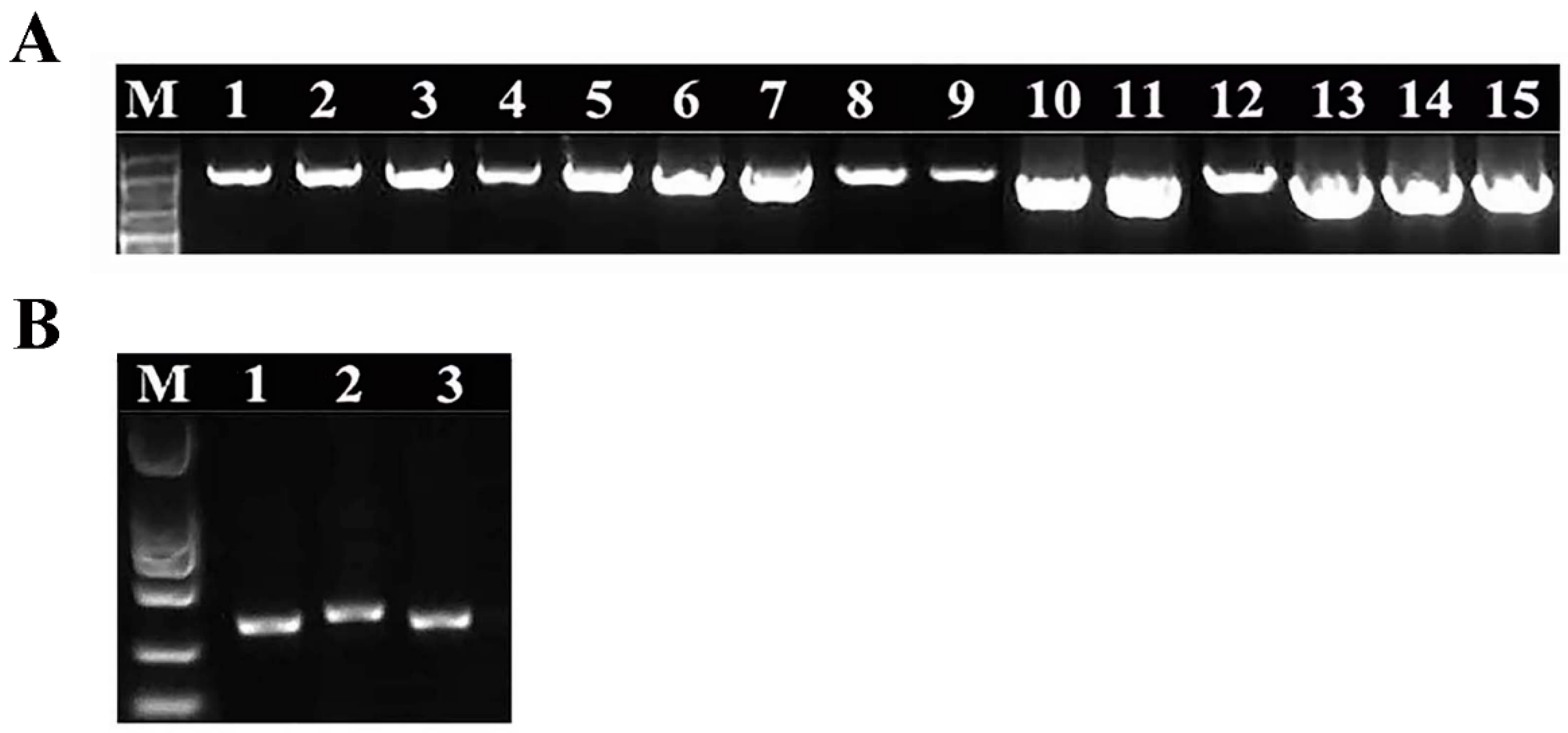
3.3. Molecular Analysis of Bacteria and Fungi

In order to clarify the evolutionary relationship among the bacteria, the phylogenetic tree was constructed based on the 16S rDNA sequences. The results showed that Bacillus siamensis, B. koreensis and B. aryabhattai B8 formed a single cluster with a close cluster of Enterobacter E24. B. amyloliquefaciens Y5 and Enterobacter Glu2 formed a single cluster, while B. amyloliquefaciens BV2007 possessed high similarity to K. michiganensis (Figure 5A).
Moreover, the phylogenic tree in fungi showed that the Peniophora sp. strain formed a single cluster with a reference strain Peniophora M161, while P. oligotrophica and C. crousii formed a single cluster, indicating a close relationship between them (Figure 5B).

3.4. Drugs Screening from Antimicrobial Susceptibility Results

Based on the susceptibility tests, 15 isolates were all resistant to 100 μg/mL of Amp and 0.0625% PS, 13 isolates were resistant to 200 μg/mL of Spc, and 7 isolates were resistant to 250 μg/mL of Ery (Table 3; Figures S1 and S2). Importantly, the bacteria were all sensitive to 50 μg/mL of Kan, except for K. michiganensis and Enterobacter Glu2, but they were both sensitive to 25 μg/mL of Chl (Table 3; Figures S1 and S2). Thus, a combination of 50 μg/mL of Kan and 25 μg/mL of Chl were selected for potential therapy of bacterial pollution in MS media.
In addition, some drugs, including nano-Ag, PS, SB and SD, were screened for the resistance of fungi. The results showed that PS could completely inhibit the three fungi growths at series concentrations of 0.0125% (w/v), 0.25% (w/v), 0.5% (w/v) and 1.0% (w/v).
In contrast, less than 0.25% SB was not free of C. crousii and 0.125% SD could no longer inhibit the three fungi production (Figure S3). No inhibitory effects were observed in nano-Ag, Spc and Chl (Figure 6). Thus, PS had remarkably inhibitory effects on the fungi and was selected for the prevention of fungal pollution in MS media.

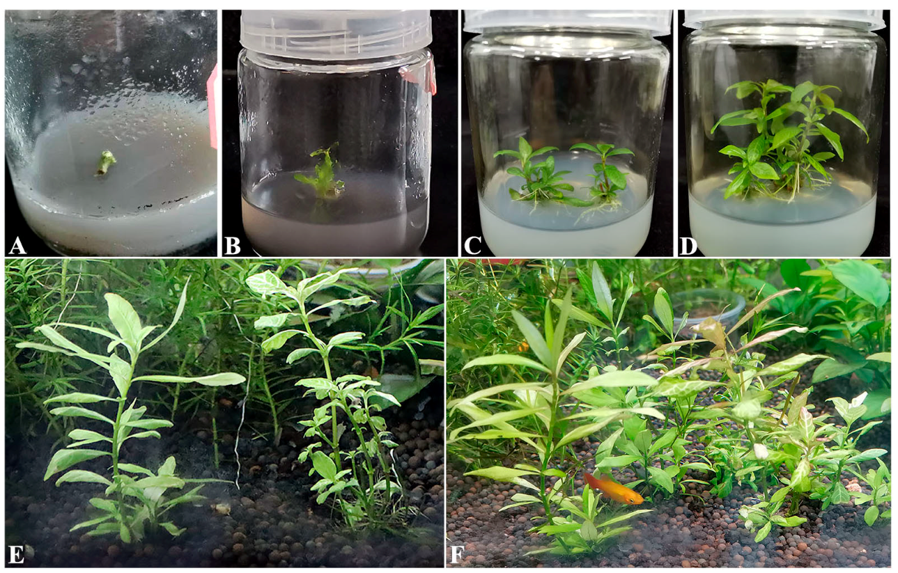
3.5. The Application of Antimicrobial Agents in Tissue Culture

To avoid the influences on the H. polysperma explants or seedlings, different concentrations of the above antimicrobial agents were tested. The results showed that a combination of 10 μg/mL of Kan and 5 μg/mL of Chl had good antibacterial activity in MS media and also no side effects on the explants’ growth, while when using lower than those concentrations, severe pollution occurred in MS media (Figure 7A–D; Figure S4).
Moreover, 0.015625% PS had good performance for antifungal activity. In contrast, higher than 0.015625% PS, from 0.03125% to 0.5%, caused worse influences on the seedlings’ growth. Similar effects were observed under the treatments of SB and SD, respectively (Figure S4; Table S1).
Hence, a combination of 10 μg/mL of Kan and 5 μg/mL of Chl, and 0.015625% PS were well applied in MS media to prevent the microbial pollution. Subsequently, H. polysperma explants were in normal progress of tissue culture, and finally the H. polysperma seedlings were transplanted and grew well in the aquarium (Figure 7E,F).

4. Discussion

Aquatic plants grew in freshwater and saltwater with varied life cycles and architectures, and their total diversity is abundant [41]. Aquatic macrophytes, belonging to multicellular species, mostly grew in freshwater. The macrophytes commonly have soft stems with branch shapes of low and creeping clumps, and exhibit in free-floating, emergent or submerged forms, such as Pistia stratiotes, Myriophyllum aquaticum, Egeria densa and Typha angustifolia. Aquatic plants could usually reproduce rapidly via vegetative means. Among them, the tissue culture was used as an important method for mass production in business. Plenty of aquatic plants had been reported, including free-floating Nymphoides indica [42] and P. stratiotes [24], rooted Anubias barteri [43], emergent Nymphaea lotus [1], submerged Rotala rotundifolia [4], Ludwigia repens [44], Cryptocoryne lucens [45,46] and C. wendtii [47]. In H. polysperma, it was previously found that multiple adventitious shoots were easily induced from the leaf explants, with the additive of 0.1 mg/L TDZ and 0.10 mg/L IBA in MS media [3]. In this study, 1.0 mg/L 6-KT and 1.0 mg/L IBA were added in MS media, and adventitious shoots were well induced from the stem explants, hinting that callus inducing from different explants was dependent on the different concentration of phytohormones.
Plants closely associated with “endophyte” living within plant tissues that are not eradicated after surface sterilization, can interfere with or inhibit the initiation and growth of in vitro cultures [48]. The endophytes usually keep symbiotic relationships with plants for mutual benefits. Accidentally, over-reproduction of endophytes caused the bacterial pollution. The endophytes from a variety of plants had been previously observed by microscopy technique via fluorescent in situ hybridization (FISH) [49,50,51,52,53] and identified by 16S rDNA sequencing [37,50,54,55]. For instance, Bacillus, Pseudomonas, Fusarium, Burkholderia, Rhizobium and Klebsiella were identified as the most dominant genera in crops, including wheat, maize, rice and bean, etc. [10]. Pantoea, Enterobacter, Agrobacterium, and Methylobacterium spp., were identified as endophytes in grape, banana, cotton, clover and potato [11,12,13,14]. Significantly, Pseudomonas and Burkholderia were found to fight against pathogens and pests as antagonists, and Klebsiella participated in nitrogen fixation [8,14,51,56]. Besides, Pseudomonas and Bacillus were found colonized on roots of plants [49], and probably migrated upward and reached different tissues of plants later [10,57]. Especially, Proteobacteria, including Pseudomonas and Enterobacter was predominantly isolated from the callus stocks of grapevine, barley, tobacco and Arabidopsis [9]. In the ornamental aquarium plants, Bacillus, Enterobacter, Klebsiella and Pseudomonas were detected early [15]. Consistently with the previous reports, Bacillus spp., Enterobacter, Pantoea and Klebsiella strains in the present study were identified from the contaminants in the MS media of H. polysperma, suggesting a common bacterial pollution among different aquatic and land plant species. Especially, Kosakonia and Ensifer strains were firstly reported in the tissue culture of H. polysperma in this study. Whether these bacteria are endophytes or not needs to be further investigated via whole-metagenome profiling (WMG), which could clarify the taxonomic and functional diversity of bacteria [8,10,50,54]. Moreover, three kinds of fungi belong to Plectosphaerella, Cladosporium and Peniophora, not previously reported in eukaryotic endophytes in plants [54], and so it was inferred that they were derived from the external working environment.
Excessive proliferation of endophytes and the microbial pollution from the external conditions would severely hinder the progress of tissue culture. Traditionally, plant material was surface-disinfected with a treatment of 70–75% ethanol, 2% NaClO, or 0.1% HgCl2. However, these disinfectants are also harmful to plant cells and needed to be used at an appropriate dose and time. In this case, certain types of microorganisms, such as Pseudomonas, Agrobacterium and Klebsiella spp. [58], are likely not to be destroyed; the endophytes might be also protected from the surface disinfection treatment. Accordingly, in this study, the H. polysperma explants were heavily infected with multiple bacteria and fungi, which occurred in different colors and shapes. In order to eliminate the unwanted microbes, several drugs were tested. As previously reported, antibiotics induced side effects on the chrysanthemum and tobacco growth in vitro [16], so the appropriate inhibitory concentrations were determined to avoid the negative influences on the explants [1]. Expectedly, a mixture of 10 μg/mL of Kan, 5 μg/mL of Chl and 0.015625% PS could inhibit the microbial infection and was well applied in the MS media of H. polysperma. Usually, the explants are not always infected at the establishment stage, but frequently this occurs later in the multiplication stage after several subcultures [1]. Thus, the medicine might be more needed at the later propagation than at the early initial stage. Recently, wide ranges of nanoparticles (NPs) were reported to possess antimicrobial activity, including NPs of silver (Ag), silicon dioxide (SiO2), titanium dioxide (TiO2), and Zinc oxide (ZnO) [1]. However, in this study, no inhibitory effect was found in nano-Ag against the fungi, possibly due to its low additive dose.
Noteworthily, a number of endophytic bacteria were favorable for promoting plant growth, associated with an increase in nutrient availability and the production of the endogenous phytohormone [49]. For instance, auxin–producing bacterial strains from Pseudomonas, Enterobacter, Rhizobium, Bradyrhizobium, Bacillus, Methylobacterium, Rhodococcus, Acinetobacter and Microbacterium had been reported to improve the endogenous IAA level of the Triticum aestivum plant [59,60]. Considering the positive effect of endophytic bacteria in plants, it might be rational to keep a balance of the total microbial community rather than completely kill them during the tissue culture [49]. In future, it might be helpful to solve this problem from the next-generation sequencing (NGS)-mediated explorations, which could uncover the microbial communities profile and provide considerable taxonomic diversity with functional implications.
In conclusion, this report revealed that the bacteria, commonly in Bacillus, Enterobacter, Pantoea and Klebsiella, and the fungi, containing Peniophora sp., P. oligotrophica and C. crousii, were reported in the contaminated MS media of H. polysperma explants. Three antimicrobial agents, 10 μg/mL of Kan, 5 μg/mL of Chl and 0.015625% PS, were screened out to be applied jointly in culture media to prevent the microbial pollution, which provided a practical reference for the modified MS media on the tissue culture of other aquatic plants.

Supplementary Materials

The following supporting information can be downloaded at: https://www.mdpi.com/article/10.3390/microorganisms10122476/s1, Figure S1: The inhibition effects of the antibiotics for bacteria in LB broth. Figure S2: The inhibition effects of chloramphenicol for bacteria in LB broth. Figure S3: The inhibition effects of three antifungal agents for fungi in PDA plates. Figure S4: The influence of two kinds of antibiotics on H. polysperma explants during tissue culture. Figure S5: The influence of three antifungal agents on H. polysperma explants during tissue culture. Figure S6: The normal growth of H. polysperma explants treated with a mixture of three antimicrobial agents in MS media. Table S1: Phenotypes of explants or seedlings in MS media treated with three antifungal agents.

Author Contributions

W.L., G.C., M.Z., Y.Z., R.Z., W.Y., Z.Z. and Y.G. performed the experiments and analyzed the data. W.L., G.C., M.Z., Y.Z., R.Z., Z.Z. and Y.G. wrote the manuscript with input from all authors. Y.S., B.Z. and J.T. contributed reagents and materials. Y.S. designed the study, and reviewed and edited the manuscript. All authors have read and agreed to the published version of the manuscript.

Funding

The work was supported by the National Natural Science Foundation of China (31902408) and the Doctoral Sustentation Fund of Qingdao Agricultural University (1120035).

Institutional Review Board Statement

Not applicable.

Informed Consent Statement

Not applicable.

Data Availability Statement

Not applicable.

Conflicts of Interest

The authors declare no conflict of interest.

References

  1. Sulaiman, E.S. Development of Sterilization Procedures and In Vitro Studies of Nymphaea lotus. Master’s Thesis, Universiti Putra Malaysia, Serdang, Malaysia, 2004. [Google Scholar]
  2. Stanly, C.; Bhatt, A.; Keng, K.L. An efficient in vitro plantlet regeneration of Cryptocoryne wendtii and Cryptocoryne becketti through shoot tip culture. Acta Physiol. Plant 2011, 33, 619–624. [Google Scholar] [CrossRef]
  3. Karataş, M.; Aasim, M.; Çınar, A.; Dogan, M. Adventitious shoot regeneration from leaf explant of dwarf hygro (Hygrophila polysperma (Roxb.) T. Anderson). Sci. World J. 2013, 2013, 680425. [Google Scholar] [CrossRef] [PubMed] [Green Version]
  4. Dogan, M. The effectiveness of light emitting diodes on shoot regeneration in vitro from shoot tip tissues of Limnophila aromatica (Lamk.) Merr. and Rotala rotundifolia (Buch-Ham. ex Roxb) Koehne. Biotech. Histochem. 2020, 95, 225–232. [Google Scholar] [CrossRef] [PubMed]
  5. Kim, D.H.; Gopal, J.; Sivanesan, I. Nanomaterials in plant tissue culture: The disclosed and undisclosed. RSC Adv. 2017, 7, 36492–36505. [Google Scholar] [CrossRef] [Green Version]
  6. Sivanesan, I.; Muthu, M.; Gopal, J.; Tasneem, S.; Kim, D.H.; Oh, J.W. A fumigation-based surface sterilization approach for plant tissue culture. Int. J. Environ. Res. Public Health 2021, 18, 2282. [Google Scholar] [CrossRef]
  7. Hardoim, P.R.; van Overbeek, L.S.; Elsas, J.D. Properties of bacterial endophytes and their proposed role in plant growth. Trends Microbiol. 2008, 16, 463–471. [Google Scholar] [CrossRef]
  8. Thomas, P.; Sekhar, A.C.; Shaik, S.P. High taxonomic diversity of cultivation-recalcitrant endophytic bacteria in grapevine field shoots, their in vitro introduction, and unsuspected persistence. Planta 2017, 246, 879–898. [Google Scholar] [CrossRef]
  9. Thomas, P.; Franco, C.M.M. Intracellular bacteria in plants: Elucidation of abundant and diverse cytoplasmic bacteria in healthy plant cells using in vitro cell and callus cultures. Microorganisms 2021, 9, 269. [Google Scholar] [CrossRef]
  10. Rana, K.L.; Kour, D.; Kaur, T.; Devi, R.; Yadav, A.N.; Yadav, N.; Dhaliwal, H.S.; Saxena, A.K. Endophytic microbes: Biodiversity, plant growth-promoting mechanisms and potential applications for agricultural sustainability. Antonie Van Leeuwenhoek 2020, 113, 1075–1107. [Google Scholar] [CrossRef]
  11. McInroy, J.A.; Kloepper, J.W. Survey of indigenous bacterial endophytes from cotton and sweet corn. Plant Soil 1995, 173, 337–342. [Google Scholar] [CrossRef]
  12. Sturz, A.V.; Christie, B.R.; Matheson, B.G. Association of bacterial endophyte populations from red clover and potato crops with potential for beneficial allelopathy. Can. J. Microbiol. 1998, 44, 162–167. [Google Scholar] [CrossRef]
  13. Habiba, U.; Reza, S.; Saha, M.L.; Khan, M.R.; Hadiuzzaman, S. Endogenous bacterial contamination during in vitro culture of banana: Identification and prevention. Plant Tissue Cult. 2002, 12, 117–124. [Google Scholar]
  14. Compant, S.; Kaplan, H.; Sessitsch, A.; Nowak, J.; Ait Barka, E.; Clément, C. Endophytic colonization of Vitis vinifera L. by Burkholderia phytofirmans strain PsJN: From the rhizosphere to inflorescence tissues. FEMS Microbiol. Ecol. 2008, 63, 84–93. [Google Scholar] [CrossRef] [PubMed] [Green Version]
  15. Trust, T.J.; Bartlett, K.H. Isolation of Pseudomonas aeruginosa and other bacterial species from ornamental aquarium plants. Appl. Environ. Microbiol. 1976, 31, 992–994. [Google Scholar] [CrossRef] [PubMed] [Green Version]
  16. Teixeira da Silva, J.A.; Nhut, D.T.; Tanaka, M.; Fukai, S. The effect of antibiotics on the in vitro growth response of chrysanthemum and tobacco stem transverse thin cell layers (tTCLs). Sci. Hortic. 2003, 97, 397–410. [Google Scholar] [CrossRef]
  17. Mantell, S.H. Microbes intimately associated with tissue and cell cultures of tropical Dioscorea yams. Plant Cell Tissue Org. 1998, 52, 47–52. [Google Scholar] [CrossRef]
  18. Stout, L.M.; Dodova, E.N.; Tyson, J.F.; Nusslein, K. Phytoprotective influence of bacteria on growth and cadmium accumulation in the aquatic plant Lemna minor. Water Res. 2010, 44, 4970–4979. [Google Scholar] [CrossRef] [Green Version]
  19. Quambusch, M.; Brümmer, J.; Haller, K.; Winkelmann, T.; Bartsch, M. Dynamics of endophytic bacteria in plant in vitro culture: Quantification of three bacterial strains in Prunus avium in different plant organs and in vitro culture phases. Plant Cell Tissue Organ 2016, 126, 305–317. [Google Scholar] [CrossRef]
  20. Bolton, J.J. What is aquatic botany and why algae are plants-the importance of non-taxonomic terms for groups of organisms. Aquat. Bot. 2016, 132, 1–4. [Google Scholar] [CrossRef]
  21. Horppila, J.; Nurminen, L. Effects of submerged macrophytes on sediment resuspension and internal phosphorus lading in Lake Hiidenvesi (southern Finland). Water Res. 2003, 37, 4468–4474. [Google Scholar] [CrossRef]
  22. Gu, C.G.; Li, F.F.; Xiao, J.B.; Chu, S.Y.; Song, S.; Wong, M.H. A novel submerged Rotala rotundifolia, its growth characteristics and remediation potential for eutrophic waters. Sci. Rep. 2019, 9, 14855. [Google Scholar] [CrossRef] [PubMed]
  23. Espinoza-Quiñones, F.R.; Zacarkim, C.E.; Palacio, S.M.; Obregόn, C.L.; Zenatti, D.C.; Galante, R.M.; Rossi, N.; Rossi, F.L.; Pereira, I.R.A.; Welter, R.A.; et al. Removal of heavy metal from polluted river water using aquatic macrophytes Salvinia sp. Braz. J. Phys. 2005, 35, 744–746. [Google Scholar] [CrossRef]
  24. Zhang, Y.; Wang, Y.; Yang, B.; Chen, S. In vitro regeneration and propagation of Pistia stratiotes: An ideal aquatic plant for biomanufacturing and bioremediation. Chin. J. Appl. Environ. Biol. 2008, 4, 445–449. [Google Scholar]
  25. Carlos, A.; Harguinteguy, M.; Pignata, L.; Fernández-Cirelli, A. Nickel, lead and zinc accumulation and performance in relation to their use in phytoremediation of macrophytes Myriophyllum aquaticum and Egeria densa. Ecol. Eng. 2015, 82, 512–516. [Google Scholar] [CrossRef]
  26. Hanks, N.A.; Caruso, J.A.; Zhang, P. Assessing Pistia stratiotes for phytoremediation of silver nanoparticles and Ag(I) contaminated waters. J. Environ. Manag. 2015, 164, 41–45. [Google Scholar] [CrossRef]
  27. Ebrahimbabaie, P.; Meeinkuirt, W.; Pichtel, J. Phytoremediation of engineered nanoparticles using aquatic plants: Mechanisms and practical feasibility. J. Environ. Sci. 2020, 93, 151–163. [Google Scholar] [CrossRef]
  28. Antunes, C.; Correia, O.; Silva, J.M.; Cruces, A.; Freitas, M.C.; Branquinho, C. Factors involved in spatiotemporal dynamics of submerged macrophytes in a portuguese coastal lagoon under mediterranean climate. Estuar. Coast. Shelf Sci. 2012, 110, 93–100. [Google Scholar] [CrossRef]
  29. Arefin, M.A.; Rashid, F.; Islam. A review of biofuel production from floating aquatic plants: An emerging source of bio-renewable energy. Biofuels Bioprod. Biorefining 2021, 15, 574–591. [Google Scholar] [CrossRef]
  30. Tan, Q.G.; Cai, X.H.; Feng, T.; Luo, X.D. Megastigmane-type compounds from Rotala rotundifolia. Chin. J. Nat. Med. 2009, 7, 187–189. [Google Scholar] [CrossRef]
  31. Kshirsagar, A.; Ingale, K.; Vyawahare, N.; Thorve, V. Hygrophila spinosa: A comprehensive review. Pharmacogn. Rev. 2010, 4, 167–171. [Google Scholar] [CrossRef] [Green Version]
  32. Swapna, M.M.; Prakashkumar, R.; Anoop, K.P.; Manju, C.N.; Rajith, N.P. A review on the medicinal and edible aspects of aquatic and wetland plants of India. J. Med. Plants Res. 2011, 5, 7163–7176. [Google Scholar] [CrossRef]
  33. Zhang, L.J.; Yeh, S.F.; Yu, Y.T.; Kuo, L.-M.Y.; Kuo, Y.H. Antioxidative flavonol glucuronides and anti-HBsAg flavonol from Rotala rotundifolia. J. Tradit. Complement Med. 2011, 1, 57–63. [Google Scholar] [CrossRef] [PubMed]
  34. Rosen, D.J. Cryptocoryne beckettii (Araceae), a new aquatic plant in Texas. Sidahora 2000, 19, 399–401. [Google Scholar]
  35. Maki, K.; Galatowitsch, S. Movement of invasive aquatic plants into Minnesota (USA) through horticultural trade. Biol. Conserv. 2004, 118, 389–396. [Google Scholar] [CrossRef]
  36. Murashige, T.; Skoog, F. A revised medium for rapid growth and bioassays with tobacco tissue cultures. Physiol. Plant. 1962, 15, 473–497. [Google Scholar] [CrossRef]
  37. Weisburg, W.G.; Barns, S.M.; Pelletier, D.A.; Lane, D.J. 16S ribosomal DNA amplification for phylogenetic study. J. Bacteriol. 1991, 173, 697–703. [Google Scholar] [CrossRef] [Green Version]
  38. Saitou, N.; Nei, M. The neighbor-joining method: A new method for reconstructing phylogenetic trees. Mol. Biol. Evol. 1987, 4, 406–425. [Google Scholar] [CrossRef]
  39. Kimura, M. A simple method for estimating evolutionary rate of base substitutions through comparative studies of nucleotide sequences. J. Mol. Evol. 1980, 16, 111–120. [Google Scholar] [CrossRef]
  40. Wu, S.M.; Liu, G.; Jin, W.H.; Xiu, P.Y.; Sun, C.M. Antibiofilm and anti-Infection of a marine bacterial exopolysaccharide against Pseudomonas aeruginosa. Front. Microbiol. 2016, 7, 102. [Google Scholar] [CrossRef] [Green Version]
  41. Lewis, M.; Thursby, G. Aquatic plants: Test species sensitivity and minimum data requirement evaluations for chemical risk assessments and aquatic life criteria development for the USA. Environ. Pollut. 2018, 238, 270–280. [Google Scholar] [CrossRef]
  42. Jenks, M.A.; Kane, M.E.; McConnell, D.B. Shoot organogenesis from petiole explants in the aquatic plant Nymphoides indica. Plant Cell Tissue Organ Cult. 2000, 63, 1–8. [Google Scholar] [CrossRef]
  43. Huang, L.; Chang, Y.; Chang, Y. Rapid in vitro multiplication of the aquatic angiosperm, Anubias barteri var undulata. Aquat. Bot. 1994, 47, 77–83. [Google Scholar] [CrossRef]
  44. Ozturk, M.; Khawar, K.M.; Atar, H.H.; Sancak, C.; Ozcan, S. In vitro micropropagation of aquarium plant Ludwigia repens. Asia Pac. J. Mol. Biol. Biotechnol. 2004, 12, 21–25. [Google Scholar]
  45. Kane, M.E.; Davis, G.L.; McConnell, D.B.; Gargiulo, J.A. In vitro propagation of Cryptocoryne wendtii. Aquat. Bot. 1999, 63, 197–202. [Google Scholar] [CrossRef]
  46. Kane, M.E.; Gilman, E.F.; Jenks, M.A.; Sheehan, T.J. Micropropagation of the aquatic plant Cryptocoryne lucens. Hort. Sci. 1990, 25, 687–689. [Google Scholar] [CrossRef]
  47. Mo, X.; Jiang, Q. Study on rapid proliferation and color appearing of tropical aquatic grass Cryptocoryne wendtii tropica. Acta Agric. Zhejiangensis 2003, 15, 91–94. [Google Scholar]
  48. Volk, G.M.; Bonnart, R.; de Oliveira, A.C.A.; Henk, A.D. Minimizing the deleterious effects of endophytes in plant shoot tip cryopreservation. Appl. Plant Sci. 2022, 10, e11489. [Google Scholar] [CrossRef]
  49. Rosenblueth, M.; Martinez-Romero, E. Bacterial endophytes and their interactions with hosts. Mol. Plant Microbe Interact. 2006, 19, 827–837. [Google Scholar] [CrossRef] [Green Version]
  50. Pirttilä, A.M.; Laukkanen, H.; Pospiech, H.; Myllylä, R.; Hohtola, A. Detection of intracellular bacteria in the buds of Scotch pine (Pinus sylvestris L.) by in situ hybridization. Appl. Environ. Microbiol. 2000, 66, 3073–3077. [Google Scholar] [CrossRef] [Green Version]
  51. Compant, S.; Clement, C.; Sessitsch, A. Plant growth-promoting bacteria in the rhizo- and endosphere of plants: Their role, colonization, mechanisms involved and prospects for utilization. Soil Biol. Biochem. 2010, 42, 669–678. [Google Scholar] [CrossRef] [Green Version]
  52. Bragina, A.; Berg, C.; Cardinale, M.; Shcherbakov, A.; Chebotar, V.; Berg, G. Sphagnum mosses harbour highly specific bacterial diversity during their whole lifecycle. ISME J. 2012, 6, 802–813. [Google Scholar] [CrossRef] [PubMed]
  53. Hardoim, P.R.; van Overbeek, L.S.; Berg, G.; Pirttilä, A.M.; Compant, S.; Campisano, A.; Döring, M.; Sessitsch, A. The hidden world within plants: Ecological and evolutionary considerations for defining functioning of microbial endophytes. Microbiol. Mol. Biol. Rev. 2015, 79, 293–320. [Google Scholar] [CrossRef] [PubMed] [Green Version]
  54. Tsoktouridis, G.; Tsiamis, G.; Koutinas, N.; Mantell, S. Molecular detection of bacteria in plant tissues, using universal 16S ribosomal DNA degenerated primers. Biotechnol. Biotechnol. Equip. 2014, 28, 583–591. [Google Scholar] [CrossRef] [PubMed]
  55. Edgar, R.C. Updating the 97% identity threshold for 16S ribosomal RNA OTUs. Bioinformatics 2018, 34, 2371–2375. [Google Scholar] [CrossRef] [PubMed] [Green Version]
  56. Berg, G.; Rybakova, D.; Grube, M.; KÖberl, M. The plant microbiome explored: Implications for experimental botany. J. Exp. Bot. 2016, 77, 995–1002. [Google Scholar] [CrossRef]
  57. Sturz, A.V.; Nowak, J. Endophytic communities of rhizobacteria and the strategies required to create yield enhancing associations with crops. Appl. Soil Ecol. 2000, 15, 183–190. [Google Scholar] [CrossRef]
  58. Leifert, C.; Cassells, A. Microbial hazards in plant tissue and cell cultures. In Vitro Cell Dev. Biol. Plant 2001, 37, 133–138. [Google Scholar] [CrossRef]
  59. Tsavkelova, E.A.; Cherdyntseva, T.A.; Netrusov, A.I. Auxin production by bacteria associated with orchid roots. Microbiology 2005, 74, 46–53. [Google Scholar] [CrossRef]
  60. Ali, B.; Sabri, A.N.; Ljung, K.; Hasnain, S. Auxin production by plant associated bacteria: Impact on endogenous IAA content and growth of Triticum aestivum L. Lett. Appl. Microbiol. 2009, 48, 542–547. [Google Scholar] [CrossRef]
Figure 1. Phenotypes of microbial contamination in tissue culture flasks. (AF), Various kinds of bacteria were grown around the explants in MS contaminated media during tissue culture. (A,G,H), The fungi of mixed species appeared in MS contaminated media during tissue culture. The red arrows indicate the bacteria and the blue arrows indicate the fungi.
Microorganisms 10 02476 g001
Figure 2. Morphological characterizations and the 16S rDNA amplification of bacteria obtained from the contaminated media. Morphology of 15 bacterial isolates grown in LB or TSB plates, respectively. (A) Kosakonia oryzendophytica strain (Ko). (B) Pantoea sp. strain BHUJPCS-26 (BH). (C) Bacillus amyloliquefaciens strain BV2007 (BV). (D) Bacillus siamensis strain (Bsi). (E) Bacillus subtilis strain (Bsu). (F) Bacillus amyloliquefaciens strain Y5 (Y5). (G) Bacillus koreensis strain (Bk). (H) Bacillus aryabhattai strain B8W22T.35 (B8). (I) Ensifer sp. strain BO-30 (BO). (J) Bacillus zanthoxyli strain (Bz). (K) Bacillus aryabhattai strain WH6 (Ba). (L) Enterobacter cloacae strain PR3 (Ec). (M) Klebsiella michiganensis strain (Km). (N) Enterobacter sp. strain Glu2 (Glu2). (O) Enterobacter sp. strain E24 (E24).
Microorganisms 10 02476 g002
Figure 3. 16S rDNA amplification of the bacteria and ITS amplification of fungi isolated from unwanted contaminants in culture media. (A) Lanes 1–15 indicated Ko, BH, BV, Bsi, Bsu, Y5, Bk, B8, BO, Bz, Ba, Ec, Km, Glu2 and E24, and DNA extracted from 15 bacterial isolates, respectively. 16S rDNA was amplified from the DNA template and strong PCR amplification fragments were produced as shown, as approximately 1600 bp long electrophoretic bands. (B) Lanes 1–3 indicated Peniophora sp. strain (Peniophora sp., Pe), Plectosphaerella oligotrophica strain (P. oligotrophica, Po) and Cladosporium crousii strain (C. crousii, Cc), and DNA extracted from three fungal isolates, respectively. ITS was amplified from the DNA template and a strong PCR amplification fragments were produced as shown approximately 400 bp long electrophoretic bands. Lane M, 2000 bp DNA ladder.
Microorganisms 10 02476 g003
Figure 4. Phenotypic characterizations of fungi obtained from the contaminated media. Morphology of three fungi grown in PDA plates, respectively. (AC) indicates the fungus Pe, Po and Cc, respectively. the morphologies of the fungi were observed by scotch cellophane tape method, which presented in (DF), respectively.
Microorganisms 10 02476 g004
Figure 5. Phylogenetic tree of the detected bacteria and fungi. Phylogenetic trees were constructed and the relationships were showed among the detected bacteria (A) and fungi (B) obtained from unwanted contaminants in culture media, respectively. The branching pattern was generated by the neighbour-joining method. Bootstrap analysis was performed with 1000 repetitions. Bar, 0.5 or 1.0 nucleotide substitutions per site. GenBank accession numbers are given in parentheses.
Microorganisms 10 02476 g005
Figure 6. Effects of antimicrobial agents on the three species of fungi. (AE) indicated the fungi Pe confronting with 0.25% PS, 1000 ppm nanoscale silver (nano-Ag), 200 μg/mL of Spc, 25 μg/mL Chl and the control (MS media), in turn. (FJ) indicated the fungi Cc and (KO) indicated the fungi Po strain treated with the same antimicrobial agents, respectively.
Microorganisms 10 02476 g006
Figure 7. The phenotypic response of H. polysperma explants to the antimicrobial agents. (A) The stem explants of H. polysperma were grown in the MS medium supplemented with 5 µg/mL of Chl, 10 µg/mL of Kan and 0.015625% PS. Multiple shoots were initiated from the explants after two weeks (B), and then the seedlings were grown under the treatment of three drugs for four weeks (C) and six weeks (D). Subsequently, the seedlings were acclimatized and transported into aquariums for one month (E) and two month (F), respectively.
Microorganisms 10 02476 g007
Table 1. The species analysis of bacteria from the contaminated media.
Bacterial NamesAbbreviationsCharacteristics
Kosakonia oryzendophytica strainKowhite
Pantoea sp. strain BHUJPCS-26BHwhite
Bacillus amyloliquefaciens strain BV2007BV wrinkled
Bacillus siamensis strainBsiwrinkled
Bacillus subtilis strainBsuMucus
Bacillus amyloliquefaciens strain Y5Y5Mucus
Bacillus koreensis strainBkMucus
Bacillus aryabhattai strain B8W22T.35B8Mucus
Ensifer sp. strain BO-30BO droplet
Bacillus zanthoxyli strainBzDry white sediment
Bacillus aryabhattai strain WH6BaDry white sediment
Enterobactercloacae strain PR3EcLight yellow
Klebsiella michiganensis strainKmYellow
Enterobacter sp. strain Glu2Glu2Yellow
Enterobacter sp. strain E24E24Yellow
Table 2. The species analysis of fungi from the contaminated media.
Fungal NamesAbbreviationsCharacteristics
Peniophora sp. strainPeWhite villus
Plectosphaerella oligotrophica strainPoTasteless white villus
Cladosporium crousii strainCcTasteless green villus
Table 3. Antimicrobial susceptibility testing of the bacterial strains isolated from the contaminated culture media.
Bacterial Name KanAmpErySpcChlPS
K. oryzendophytica+++++
Pantoea BH+++++
B. amyloliquefaciens BV++++
B. siamensis+++
B. subtilis+++
B. amyloliquefaciens Y5++
B. koreensis+++
B. aryabhattai B8++
Ensifer BO ++++
B. zanthoxyli+
B. aryabhattai WH6+++
E. cloacae+++
K. michiganensis+++++
Enterobacter Glu2+++++
Enterobacter E24+++++
Note: “+” indicates that a bacterial strain could not be inhibited by the antibiotic; “−” means that a bacterial strain could be inhibited by the antibiotic. Kan, 50 μg/mL of kanamycin. Amp, 100 μg/mL of ampicillin. Ery, 250 μg/mL of erythromycin. Spc, 200 μg/mL of spectinomycin. Chl, 25 μg/mL of chloramphenicol. PS, 0.0625% potassium sorbate.
Publisher’s Note: MDPI stays neutral with regard to jurisdictional claims in published maps and institutional affiliations.

