
The Macroscopic Structure of Wood

1
Department of Agricultural, Forestry and Food Sciences, University of Torino, Largo Paolo Braccini, 2, 10095 Grugliasco, Italy
2
Forest Biometrics Laboratory, Faculty of Forestry, “Stefan cel Mare” University of Suceava, Str. Universitatii 13, 720229 Suceava, Romania
*
Author to whom correspondence should be addressed.
Forests 2023, 14(3), 644; https://doi.org/10.3390/f14030644
Submission received: 7 February 2023
/
Revised: 9 March 2023
/
Accepted: 15 March 2023
/
Published: 21 March 2023
(This article belongs to the Special Issue Reviews on Structure and Physical and Mechanical Properties of Wood)
Abstract
:Understanding the macroscopic structure of wood and its formation is essential to identifying wood and evaluating its properties and quality. Depending on genetic background, environmental conditions, and tree developmental stage, the macroscopic structure of wood can vary greatly and produce specific macroscopic signatures. Here, a comprehensive outline of the wood’s macroscopic structure and the features that can be used to identify wood by macroscopic examination is presented. The planes of observations are first depicted, and the fundamental differences between softwoods and hardwoods are outlined. Then, all the different cell characteristics, arrangements, and distributions that can be macroscopically observed are illustrated with their influence on wood figure and texture and non-anatomical features.
1. Introduction
When we handle a piece of wood, we observe its esthetic characteristics and feel some of its physical properties. What we see and feel results from a highly organized agglomerate of diverse cells and the chemical compounds deposited in their lumina or on their cell walls [1]. Although wood cell types are only a few, their size, arrangement, grouping, wall thickness, and content account for wood’s remarkable aesthetic, physical, and mechanical properties [2,3,4]. The wood’s cellular origin is also originating anomalies or specific wood characteristics [5].
Based on wood features visible by the naked eye or with a 10–15× magnification lens, a trained individual can identify an unknown piece of wood or validate a supposed taxa attribution. This process is known as macroscopic wood identification (MWI). The first to realize that wood looked different in its anatomical and macroscopic structure was Anton van Leeuwenhoek (1632–1723) [6], and the first MWI atlases date back to the beginning of the 20th century [4].
MWI is based on both wood anatomical characters, defined by the cellular structure of wood, and non-anatomical ones, determined by physical, chemical, or other wood characteristics. MWI is recognized as a reliable scientific method for wood identification [7] and is extensively adopted worldwide for various applications. Being a fast, cheap, and easy-to-apply method that does not require extensive wood anatomy knowledge, MWI is particularly interesting as a field tool for forensic wood identification. It represents an effective method for the first screening in applying international regulations such as the Lacey Act Amendment in the United States [8] and the European Regulation n. 995/2010, known as European Timber Regulation (EUTR) [9]. Many references [10,11,12,13] also testify to its usefulness in the first screening of CITES-listed woods.
More recently, several authors explored the potential of automating the process of MWI through the adoption of artificial neural networks [14]. This is of particular interest in the forensic field, where various factors currently hamper MWI: training of law enforcement officers is time-consuming, wood anatomy experts able to provide such training are scarce [14], and it is not so unlikely that trained officers can be changed of duty over time, leading to a relevant dispersion of resources.
This paper aims to describe and depict the macroscopic structure of wood based on its cellular foundation after summarizing the main references of traditional and computer vision MWI. This work provides a comprehensive guide to MWI by presenting state-of-the-art descriptions and images of each character, discussing their relevance for identification and providing hints and guidelines for their interpretation and use.
The authors directly acquired magnified transverse images with XyloScope [15], a digital wood imaging device developed by the Forest Products Laboratory of Madison, Wisconsin. All images in this paper have the same magnification, and each image represents a 6.35 × 6.35 mm area.
2. Traditional and Computer Vision Macroscopic Wood Identification References
As noted by the authors in [4], despite the century-long history and the wide number of references on MWI, variability in the number and nature of diagnostic characters used by different authors is relatively small. Still, their definitions and interpretation are often varying. For this reason, the authors in [4] standardized the characters and terminology used for traditional MWI. In the following text, macroscopic characters follow the terms and definitions provided by [4] as shown in Table 1.
To provide a general overview, Table 2 reports a selection of the most recent and representative references available for both traditional and artificial intelligence MWI. A proper comparison of the advantages and disadvantages of the two methods can be hardly provided without an in-depth analysis of computer vision-based systems. Deep learning, in fact, includes different techniques such as artificial neural networks, deep neural networks, recurrent neural networks, deep reinforcement learning, and convolutional neural networks, each with its own pros and cons when applied to wood. Additionally, there is great variability in the accuracy and reliability of the currently available systems. This topic is thoroughly analyzed in [14].
3. Wood Planes of Observation
For whatever purpose, and at any magnification, identifying wood requires adequately orientating it according to three anatomical planes of reference, dictated by its original position within the stem. These are the named transverse, longitudinal radial, and longitudinal tangential (Figure 1). The transverse and longitudinal planes are respectively perpendicular and parallel to the stem longitudinal axis. The transverse plane is the one across which a tree is cut when felled by a logger; the longitudinal planes are the ones along which a board is cut from a log in a sawmill. Taking the circumference described by the stem as a reference, a radial longitudinal plane is cut along the direction of a ray (see Figure 1). In contrast, a tangential longitudinal one is cut perpendicular to the direction of a ray.
4. Fundamental Differences between the Macroscopic Structure of Softwoods and Hardwoods
The macroscopic differences between softwoods and hardwoods are related to the different cell types among the two groups and the consequent appearance of their surfaces (Table 3).
Most of the wood in conifers consists of axial tracheids [1,35]. Parenchyma cells occupy a smaller fraction of conifer woods’ volume and are organized in the radial tissues (parenchyma rays) and, in a few species, in axial strands. Earlywood tracheids, being wider and thinner-walled compared to latewood tracheids, appear lighter in color in transverse and longitudinal sections. Even if of variable width, depending on the species and year-to-year variation of growing conditions, the latewood always appears darker in color [3]. Parenchyma rays in conifers are uniseriate and therefore not visible to the naked eye on transverse and tangential sections. However, on radial surfaces, especially those obtained by splitting—not sawing—the wood, parenchyma rays might be visible as linear ribbon-like shining structures. Axial parenchyma, either scattered or tangentially oriented, is hardly visible to the naked eye but detectable under a low magnification lens.
The wood of hardwoods is more heterogeneous and consists of vessels, fibers, fiber tracheids s.l., and axial and radial parenchyma [1,36]. Vessels are circular, oval to polygonal in the transverse section, and are differently organized within the ring boundaries. Fibers usually appear darker in color compared to the other cell types as seen in the transverse section, whereas fiber tracheids and axial parenchyma usually appear lighter-colored [3]. The appearance of rays varies with their width. Wide rays are visible in all observation planes, but smaller rays might be visible only on one surface. As a rule of thumb, only rays larger than 6–8 cells are visible to the naked eye on all the planes of observation, however, the overall color of the wood also influences their appearance.
5. Wood Color in Heartwood and Sapwood
Looking at a transverse section of a log, the wood of most recent formation lies on the outside, originating from underneath the bark, while older wood lies closer to the pith (i.e., the center of the stem). As the outermost wood still functions as a means of water and solute upward transport, the innermost wood has lost this function in old enough trees. The outermost and younger wood is called sapwood, while the heartwood is the innermost and older part of a stem. Sapwood is typically yellowish and light-colored, while heartwood can be either of the same shade as sapwood (e.g., Acer spp., Abies spp., Ochroma pyramidale, etc.) or darker (e.g., Quercus spp., Larix spp., Millettia spp., etc.), and therefore easily distinguishable from it (Figure 2).
The occurrence of extractives defines heartwood color and plays a crucial role in determining the aesthetic value of timber. While woods with brown, white, and gray hues are common, red, yellow, black, purple, pink, green, orange, and variegated ones are less common to rare (Table 4 and Figure 2). During MWI, cautiousness must be paid when evaluating wood color (Table 1, features 59 to 64) because it might have been altered by different factors: color can change from green to dry wood state, moreover, dry wood color is often subject to changes resulting from exposition to light and/or contact with oxygen (air) [3]; treatments can be applied to modify it, for instance, to minimize the difference between sapwood and heartwood, or to confer valuable tones [37,38].
6. Texture
The diameter of vessels and tracheids determines wood texture in hardwoods and softwoods, respectively. Vessel elements are tube-shaped cells with a diameter ranging from less than 0.05 mm (e.g., Buxus sempervirens) to over 0.5 mm (e.g., Cedrelinga catenaeformis) when the wood is sampled in the outer wood at the base of the stem of tall plants [39,40]. Vessel diameter is macroscopically divided into three classes (Table 1, features 19 to 21). However, the distinction between two close classes (small and medium or medium and large) may be difficult depending on the sample. In such cases, while performing an MWI, it is advisable to proceed by exclusion by ruling out only the class not included in the two possible ones.
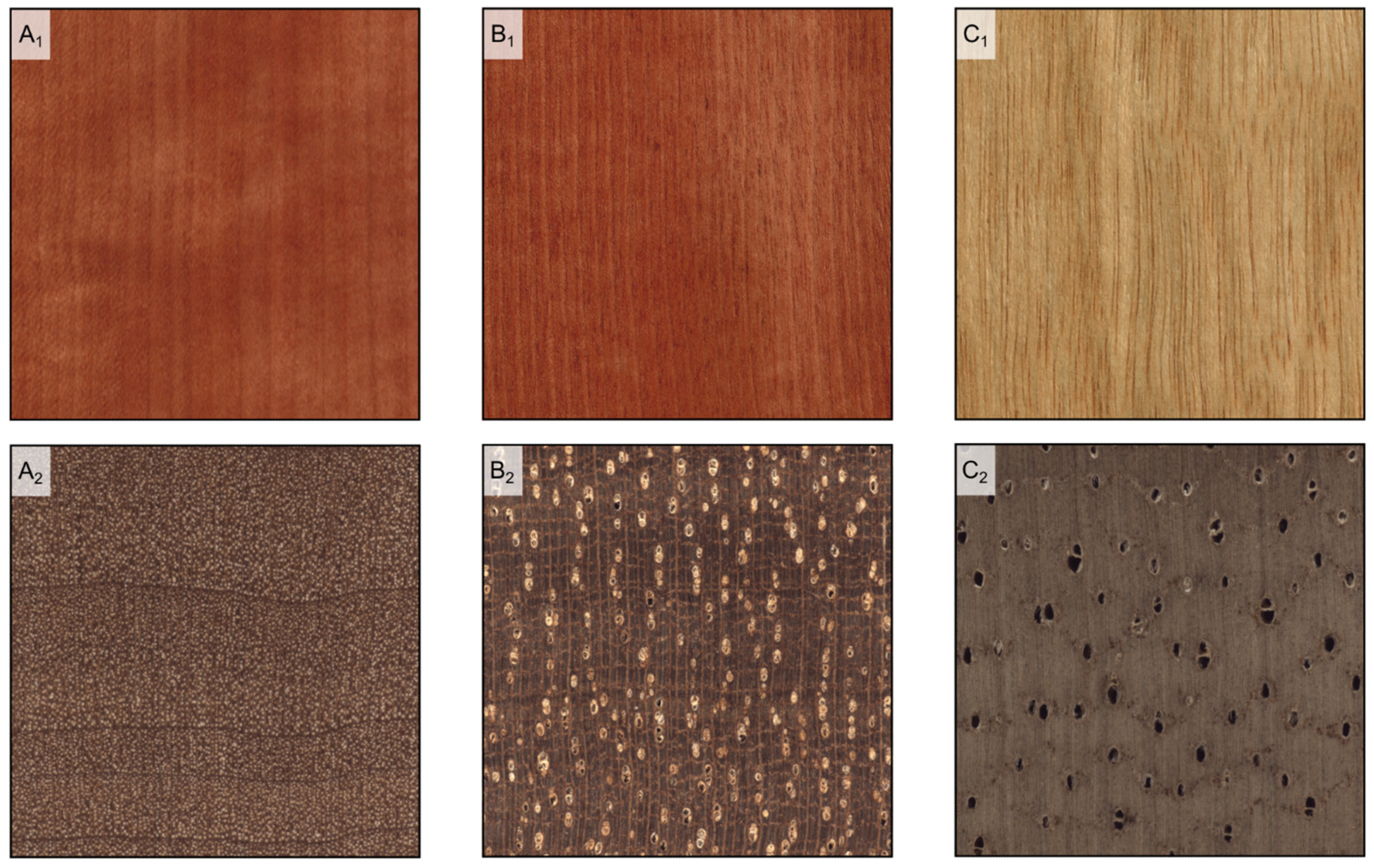
A hardwood wood texture (Table 5) is defined as fine if vessels are too narrow to be visible to the naked eye (Table 1, feature 19), medium if vessels are barely visible (Table 1, feature 20), and coarse if vessels are visible (Table 1, feature 21) (Figure 3).
In softwoods, tracheids diameter is, on average, between 0.03 and 0.06 mm. Therefore, tracheids are not visible to the naked eye.
7. Wood Figure
The term ‘figure’ in wood refers to any distinctive pattern, design, or appearance visible on a longitudinal wood surface. The figure is determined by many anatomical structures, which are presented below.
7.1. Growth Rings
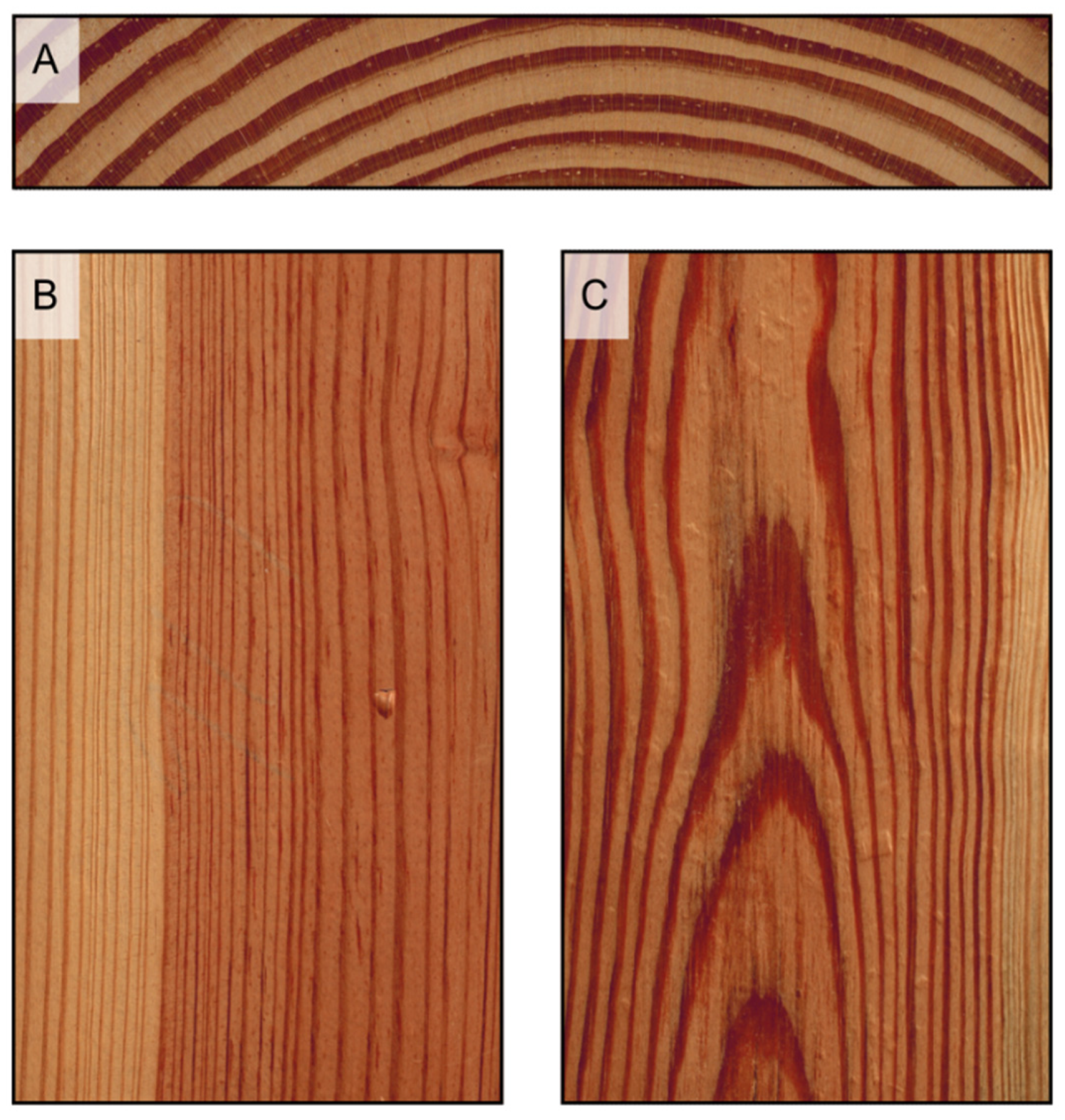
Each growing season, woody plants produce a new growth ring. Growth ring boundaries (Table 1, features 1 and 2) are visible to the naked eye on transverse surfaces as concentric circles in most temperate woods and several tropical ones. Growth rings are essential in defining the figure of wood since they draw hyperbola branches, or flames, on longitudinal tangential surfaces and parallel bands on radial surfaces (Figure 4), which are more evident the more pronounced the growth rings are. Growth rings are composed of earlywood and latewood: the more marked the transition between such portions, and the clearer the appearance of growth rings.
Growth rings are almost always distinct and visible in softwoods, while they can be indistinct in many tropical hardwoods. Caution should be paid when using growth rings for identification since their presence can be variable in several tropical hardwoods [41,42], and they can be difficult to detect macroscopically in numerous temperate ones (Table 6). Additionally, wide bands of parenchyma (e.g., Millettia spp.) and seemingly marginal bands of parenchyma (e.g., Swietenia spp.) (see “Section 7.2”) can be misinterpreted as growth ring boundaries.
The transition from earlywood to latewood can be described as abrupt or gradual in softwoods (Table 1, feature 56), with the latter being the most common one, and is marked by an increased tracheid wall thickness and smaller cell radial diameter. Tracheids are thin-walled with a wide lumen in earlywood and thick-walled with a smaller lumen in latewood. If the transition is abrupt, two distinct bands are macroscopically visible in the transverse section: a light-colored one corresponding to earlywood, and a dark-colored one corresponding to latewood. If the transition is gradual, the light-colored band blends into the dark-colored one (Figure 5). Growth rings and their consequent figures are usually more evident in woods with an abrupt transition.
Caution must be taken when using this feature for identification since many species have only one type of transition, either abrupt or gradual, but some others can present both of them (variable) depending on the growth ring (Table 7).
In hardwoods, the transition can be abrupt, gradual, or absent and is marked by porosity (Table 1, features 3 to 5), which is the difference in the diameter and density of vessel elements between earlywood and latewood. The transition is abrupt in ringporous woods, gradual in semi-ring porous woods, and absent in diffuse porous woods (Table 8).
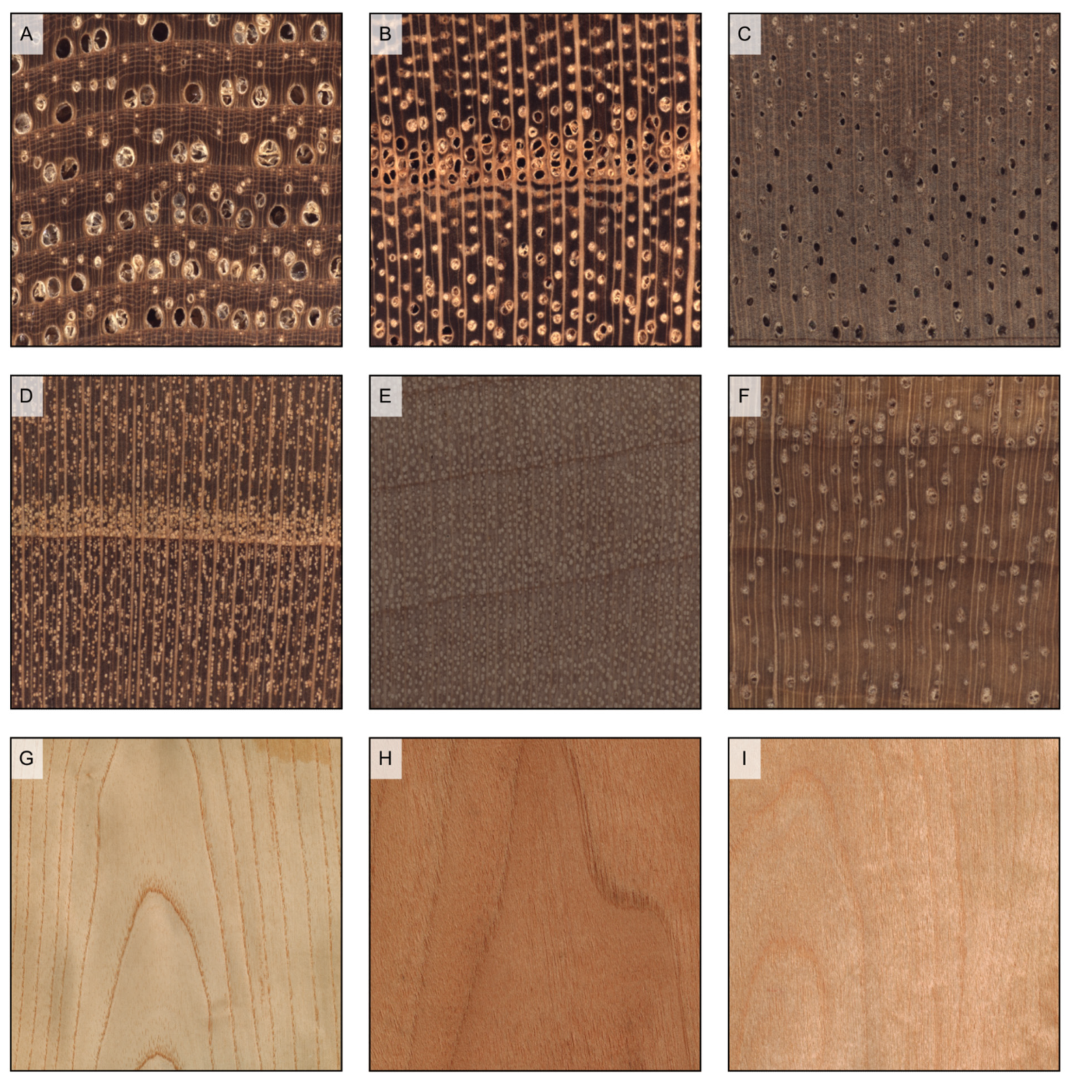
A wood is ring-porous if earlywood vessels are distinctly larger than those in the latewood of the previous and same growth ring, and form a well-defined zone or ring, clearly discernible to the naked eye by its coarser texture (Figure 6). A finer analysis of the vessels of ring-porous woods’ earlywood can discriminate between species that present one row only (e.g., Carya ovata) or multiple ones (e.g., Castanea sativa) of earlywood vessels (Table 1, feature 6). Additionally, earlywood vessels on the same row can be packed together (widest tangential spacing less than one earlywood vessel, e.g., Gleditsia triacanthos) or be farther apart (widest tangential spacing more than one earlywood vessel, e.g., Paulownia tomentosa) (Table 1, feature 7). In ring-porous woods, the figure consequent to growth rings is usually evident (Figure 6).
Earlywood vessels’ diameter gradually narrows from earlywood to latewood (e.g., Juglans regia) in a semi-ring porous wood, or earlywood vessels are of the same diameter as latewood ones but much more closely spaced (e.g., Prunus avium) (Figure 6). To the naked eye, in the first case, it is still possible to see a difference in texture between earlywood and latewood, but without a clear boundary. In the second case, earlywood appears more porous than latewood. In semi-ring porous woods, the figure consequent to growth rings is visible but not evident (Figure 6).
Vessels have more or less the same diameter and density through earlywood and latewood in diffuse porous woods (Figure 6). Therefore, the two parts are indistinguishable both to the naked eye and at the microscope. It is the most common porosity type, as almost all tropical and most temperate species are diffuse-porous. In this case, growth rings can still be marked by other structural changes, such as the presence of marginal parenchyma (see “Section 7.2”) or of thick-walled radially flattened latewood fibers (see “density”) or of distinct fiber zones close to the ring boundary (see [43] for examples). These structural changes appear to the naked eye as a darker line that delimits two adjacent growth rings. In diffuse porous woods, the figure consequent to growth rings is usually faint or barely visible to not visible (Figure 6).
While most species have only one type of porosity, in some it may vary depending on the ring or the sample. Some examples are Dalbergia cearensis, Populus spp., and Salix spp., which can vary between diffuse porous and semi-ring porous, and Cedrela odorata, Tectona grandis, and Toona hexandra, which can vary between ring-porous and semi-ring porous.
7.2. Axial Parenchyma
Wood axial parenchyma cells are brick-shaped cells, alive in sapwood, and dead in the heartwood. They have the main axis vertically oriented, are stacked one upon each other in strands, and can be distributed according to many different patterns as seen in the transverse section.
In softwoods, they are usually so scarcely diffuse to be hardly visible to the naked eye, and therefore they do not contribute to determining any figure. However, in softwoods, axial parenchyma can be detected with a loupe as tiny dark spots on the transverse section and therefore be of help in MWI (Table 1, feature 58) (Table 9 and Figure 7). Be aware that resin spots can be easily mistaken for axial parenchyma, thus possibly deceiving the identifier.
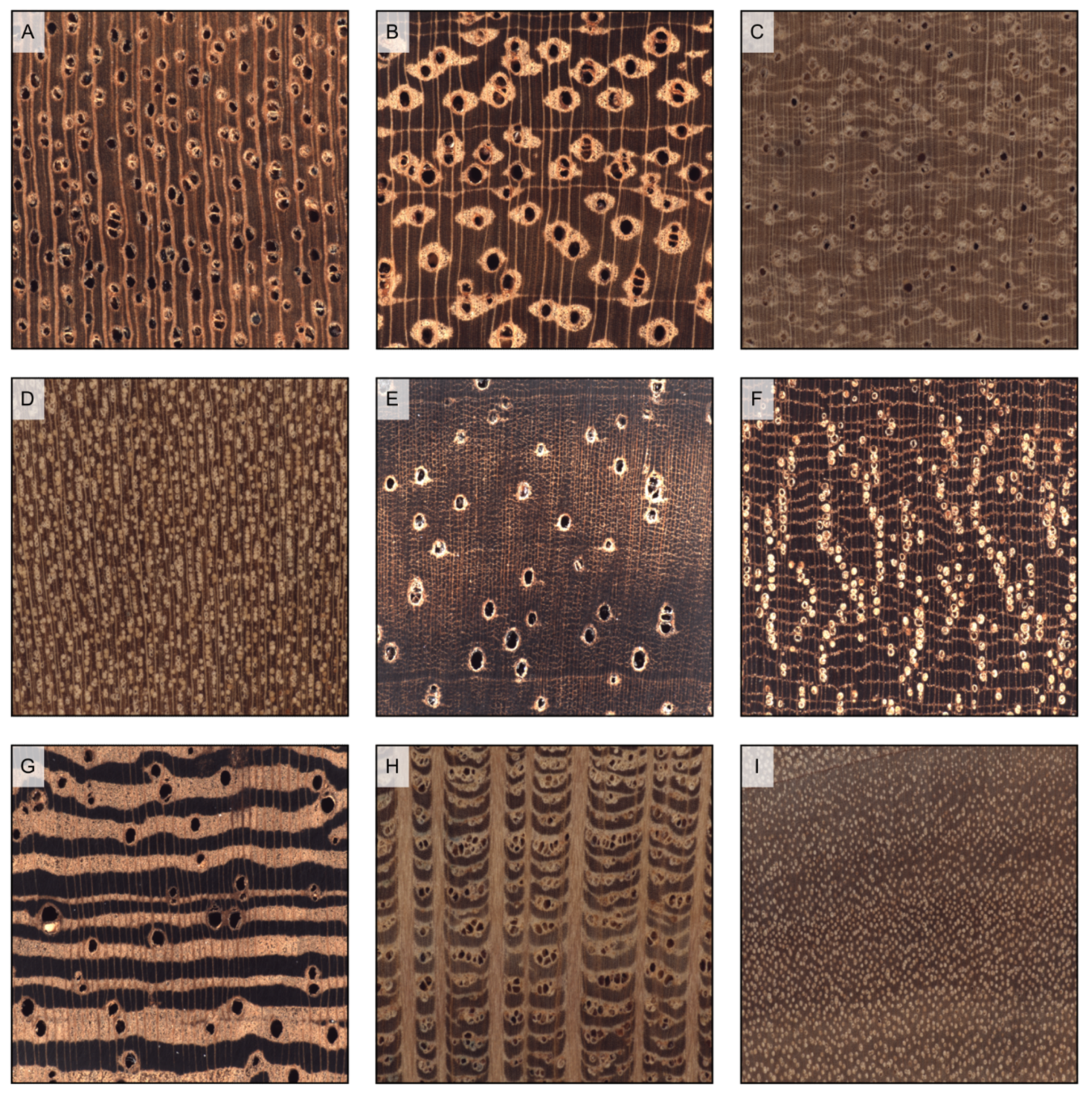
When large enough, axial parenchyma aggregates are visible to the naked eye in hardwoods as lighter-colored areas on the transverse and longitudinal planes. In hardwoods, axial parenchyma can be distributed around vessels (paratracheal) or independently from them (apotracheal). Paratracheal distributions can be vasicentric, lozenge-aliform, and winged-aliform (Table 1, features 31 to 33). The type of distribution can be determined on a transverse plane, where the parenchyma around vessels appears as a round halo in the first case, like a diamond-shaped halo in the second case, and as a halo with narrow, elongated tangential extensions in the third case (Figure 8). When the parenchyma of two or more vessels merges, it is called confluent (Table 1, feature 34). If prominent enough, paratracheal parenchyma slightly contributes to the wood figure since it appears on longitudinal surfaces as a light-colored band along the vessels’ grooves (Figure 9). Apotracheal distributions range from single cells or short discontinuous lines of cells scattered among fibers (diffuse and diffuse-in-aggregates parenchyma) to more or less pronounced tangential bands (Table 1, features 29, 30, and 35 to 41) (Figure 8). When parenchyma bands are large enough to be visible to the naked eye, they appear as flames on longitudinal tangential surfaces and parallel bands on radial surfaces, like growth rings do (Figure 9). When parenchyma bands are particularly prominent, such as in Millettia spp., they can be mistaken for growth rings.
Axial parenchyma characterizes the transverse section pattern of numerous hardwoods, and is usually more abundant in tropical ones, thus proving to be very useful in the MWI of several woods (Table 10). While the more prominent forms of distribution (e.g., large bands or lozenges around big vessels) are easily visible to the naked eye, the more faint ones (e.g., diffuse or diffuse-in-aggregates) can be hardly distinguishable even with the aid of a loupe. Usually, a drop of water on the transverse surface greatly improves the visibility of axial parenchyma. Several woods have more than one type of distribution, while in some others, axial parenchyma is absent or rare.
7.3. Radial Parenchyma (Rays)
Radial parenchyma is composed of parenchyma cells grouped in band-like structures called rays. Rays are like walls oriented along the stem radial plane, usually several cm long, a few µm to a few mm wide, and 0.01 mm to several cm high.
Softwood rays are too narrow to be visible to the naked eye on transverse and longitudinal tangential planes. In contrast, they can be seen as faint horizontal stripes on longitudinal radial ones (Figure 10). Rays are, therefore, not useful for the MWI of softwoods.
Hardwoods rays’ dimension, instead, varies depending on the species. When large enough, other than being visible to the naked eye on the transverse plane as radial lines (Table 1, feature 43), they contribute to determining the wood figure [3]. In fact, rays appear as vertical lines on the longitudinal tangential plane (Table 1, features 44 and 48) and as horizontal stripes on the longitudinal radial. As in softwoods, they can still be seen on the longitudinal radial plane even when very narrow (Figure 10).
In hardwoods, rays can be storied, i.e., arranged in more or less regular horizontal tiers as viewed on the tangential plane (Table 1, feature 47). In such cases, they are always too small to be singularly visible to the naked eye but can confer fine horizontal striations, or ripple marks, on tangential surfaces (Figure 11). Storying is considered fine if there are 4 rows or more per axial millimeter, and coarse if the rows are 3 or fewer.
While in most tropical hardwoods, rays are not or barely visible, the occurrence of big rays easily visible on both transverse and tangential surfaces is much higher in temperate ones. On the contrary, storied rays are, with few exceptions, almost only present in tropical hardwoods (Table 11). As noted for axial parenchyma, a drop of water improves the visibility of tiny rays.
On the transverse surface, with the aid of a reference grid, the number of rays per mm (Table 1, feature 49) can be measured, as well as the ratio of ray width to pore diameter (Table 1, feature 45). In some species (such as Fagus sylvatica and Platanus occidentalis), rays are slightly swollen in correspondence with the growth-ring boundary (Table 1, feature 46), a character that can be seen with the aid of a loupe only (Figure 10).
7.4. Vessels’ Arrangement, Grouping, and Frequency
The arrangement of vessels usually does not impact wood figure, except when they are in wavy tangential bands, which can determine a characteristic jagged pattern on longitudinal tangential surfaces (e.g., Ulmus spp.) (Table 1, feature 8) (Figure 12). Other vessels’ arrangements include radial, diagonal, and dendritic patterns (Table 1, features 9 to 11). The presence of specific patterns in vessels’ arrangement is not very common, and thus not useful in MWI (Table 12). More than one arrangement can be present in the same species.
Vessels’ groupings do not contribute to determining wood figure, but are useful in MWI. They can be observed on the transverse surface with the aid of a loupe and can be: exclusively solitary (90% or more), in radial multiples of four or more, in clusters, and, most commonly, solitary and in radial multiples of 2 to 3 (Table 1, features 12 to 15) (Table 13 and Figure 13). Vessels’ grouping can be difficult to be accurately evaluated when vessels are small.
A particular type of “exclusively solitary vessels” grouping is “latewood pores large, individually distinct, and few enough that they can be readily counted” (Table 1, feature 22). This character is mainly used for the separation of the red oak group (which presents the character) from the white oak one (where the character is absent instead).
As noted for vessels’ groupings, also vessels’ frequency, i.e., the number of vessels per square mm, does not contribute to determining wood figure, but is a useful character for MWI (Table 1, features 16 to 18). Vessels’ frequency (Table 14) is applied in diffuse porous and semi-ring porous woods only and must be measured on the transverse plane, more accurately with the aid of a transparent reference grid. In some species (e.g., Celtis gomphophylla and Terminalia ivorensis), vesselless tangential bands can be present (Table 1, feature 23).
7.5. Grain
Grain refers to the longitudinal alignment or pattern of axial wood cells compared to the longitudinal axis. A straight-grained wood is one in which the longitudinal cells are aligned parallel to the axis of the log or timber and is the most common case.
Several grain deviations are responsible for highly appreciated decorative figures in wood (Figure 14). Interlocked grain occurs when axial elements are aligned at an angle to the vertical axis and such spirals reverse at intervals. This produces a ribbon stripe figure on longitudinal surfaces, which is particularly pronounced on quartersawn surfaces. If axial elements have instead a wavy arrangement along the vertical axis, the grain is defined as ‘wavy’ and the consequent figure, characterized by a horizontal pattern perpendicular to the grain, is called curly or fiddleback. A less common and extremely priced figure due to a peculiar wavy arrangement of axial elements is the quilt, characterized by seemingly bubbles on longitudinal surfaces. Burls are instead the consequence of an anomalous concentration of dormant buds that force axial cells to grow in a twisted direction, giving origin to highly decorative irregular figures. Finally, crotches form in correspondence of large tree forks.
Although these alterations of grain direction are usually characteristic of certain species (Table 15), their occurrence is occasional to rare depending on the alteration type and species. They are generally not useful in MWI, except for interlocked grain, that in some species is consistent enough to be considered an identification character (Table 1, feature 105).
On the one hand, these irregular grain patterns give distinctive and highly priced wood figures, but on the other hand they are usually responsible for lower mechanical performances, anomalous shrinkage, and processing difficulties [44]. This is the case also with any other grain deviation that does not produce any decorative pattern, the most common ones being spiral grain, where axial elements are aligned at an angle to the vertical axis, and deviations due to knots, where axial cells are diverted in the proximity of a branch, whose cells are, in turn, oriented perpendicular to the grain of the trunk grain. (Figure 15).
8. Superficial Marks
During heartwood formation processes, gums and tyloses may be deposited in vessel lumina in some species (Table 16). Vessel deposits (Table 1, features 25 to 28) include a wide variety of variously colored (white, yellow, red, brown, black) chemical compounds. They can be seen as spots on transverse surfaces and frequently determine colored streaks on longitudinal ones. Tyloses (Table 1, feature 24) are balloon-like structures that, being light-reflective, can be seen shining in the vessels on the transverse plane (Figure 16).
Intercellular canals may produce distinct marks on the wood surface, too. They are tube-shaped intercellular spaces, axially or radially oriented, found in both hardwoods and softwoods; they contain resin in the latter and several types of chemical compounds in the former. Radial canals are always included in rays and are usually extremely difficult to spot macroscopically both in softwoods and hardwoods.
Axial canals in softwoods (Table 1, feature 57) are usually scattered and can be seen on the transverse surface as tiny spots darker- or lighter-colored than the background tissue. The only softwood genera that present axial canals are Larix, Picea, Pseudotsuga, and Pinus. In the first three, they are small and difficult to see even with a loupe, and in such cases, a drop of water can enhance their visibility. In Pinus, they are big enough to be often visible as streaks on longitudinal surfaces, too (Figure 17). In softwoods, the presence of radial canals is restricted to the same genera that present axial canals.
Axial canals in hardwoods (Table 1, feature 52) can be found in some tropical species, with distributions that can vary from diffuse to short or long tangential lines (Table 17).
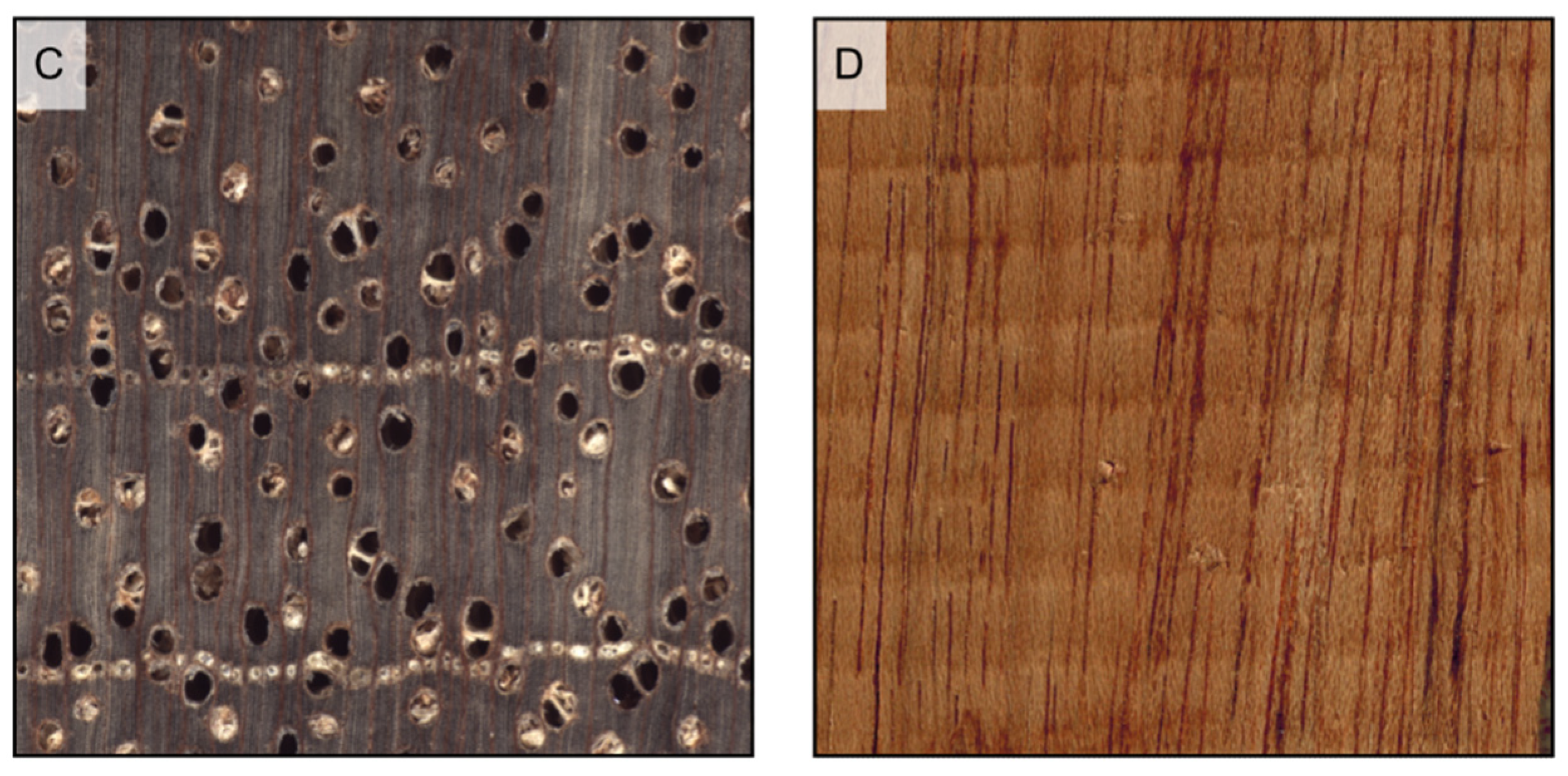
Axial canals in hardwoods can be identified on the transverse plane by their arrangement and/or by the presence of exudations, and they sometimes appear as streaks on longitudinal surfaces (Figure 18). In hardwoods, radial canals (Table 1, feature 54) are also found in tropical species, such as Antrocaryon spp., Astronium spp., Gluta renghas, and Mammea africana, but they are very hard to spot even with the aid of a loupe.
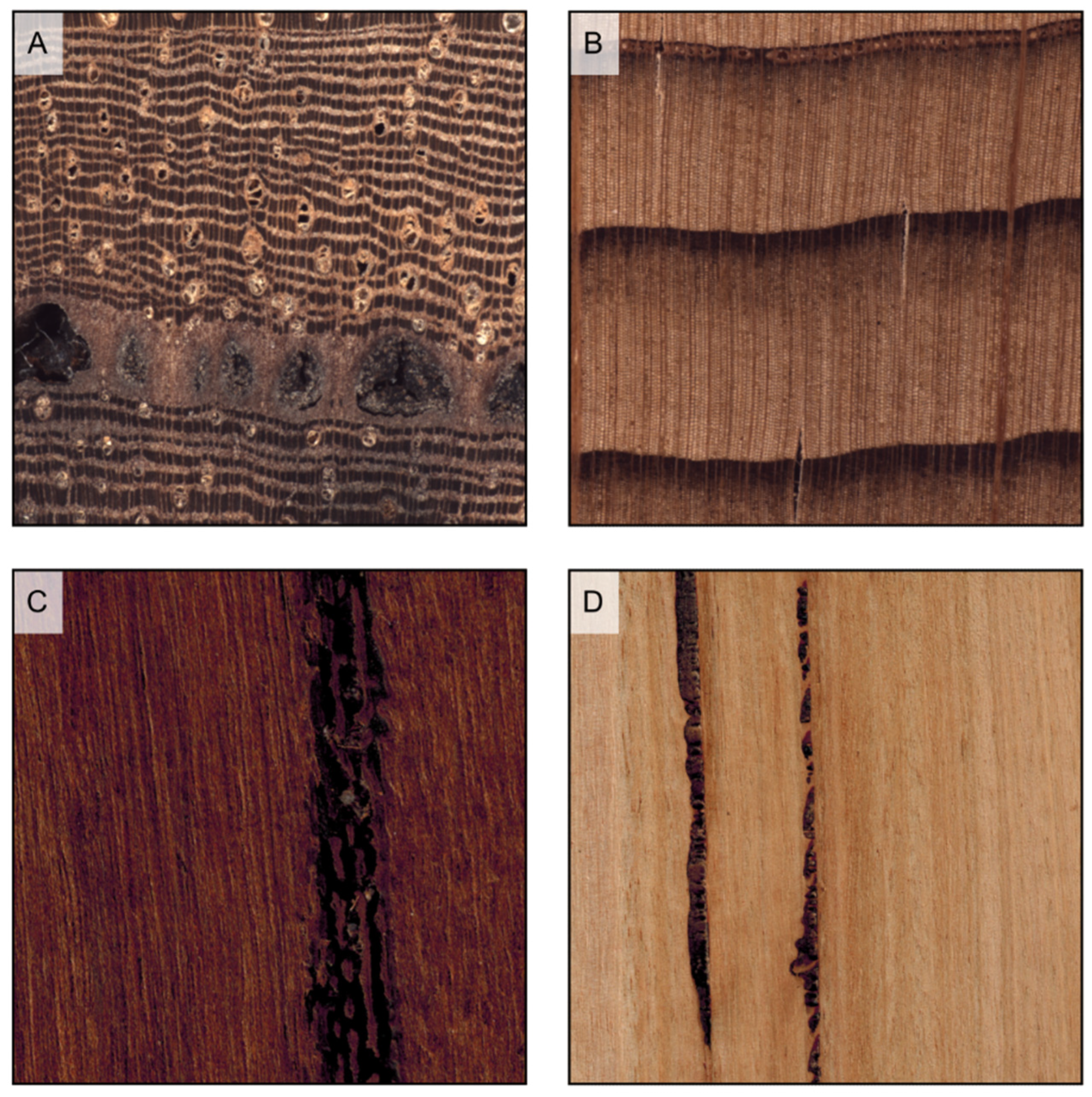
Some hardwoods and softwoods that do not have regular canals may still form so-called traumatic canals in response to traumatic events (Table 1, features 53 and 57b). They usually have an irregular outline and, being mostly larger than normal canals, are visible to the naked eye on both transverse and longitudinal planes, sometimes with the appearance of ‘scars’. This is, for instance, the case of several Eucalyptus spp., in which such traumatic canals are called ‘kino veins’ from the name of the gum they are filled with. Other examples of species that can present traumatic canals are Balfourodendron riedelianium, Cariniana micrantha, Cedrus spp., Erisma uncinatum, and Lovoa trichilioides (Figure 19).
Another possible source of superficial marks is the presence of included phloem (Table 1, feature 55), a quite uncommon feature in trees, that appears as diffuse (e.g., Aquilaria spp.) or concentric (e.g., Koompassia malaccensis) patches of bark included within the wood. Finally, some other marks of different origins may characterize the surfaces of some species (Table 1, feature 106), some of the most common ones being the pith flecks of Prunus serotina and Betula pendula (Figure 20).
9. Wood Density
Wood density (Table 1, feature 65) refers to the amount of actual wood substance (i.e., cell walls) present in a unit volume of wood, and is usually reported at a moisture content of 12%. It is a fundamental property, key for many wood end applications. With values that can range from <200 kg/m3 (e.g., Ochroma pyramidale) to >1200 kg/m3 (e.g., Dalbergia melanoxylon), its variability is much higher in hardwoods than in softwoods, where it ranges between 350 and 700 kg/m3 (Table 18).
10. Odor and Oily Surface
The presence of volatile heartwood extractives determines odor (Table 1, feature 66) that, depending on the wood, can be pleasant or unpleasant (Table 19). In several woods, the fragrance is quite characteristic and therefore very useful for identification, however, while it can be distinctively detected during wood processing, it usually fades away with wood aging.
The wood of some species, such as Tectona grandis, contains oily compounds which give a greasy feel to wood surfaces (Table 1, feature 67).
11. Heartwood Fluorescence and Other Extractives Properties
The heartwood of several hardwoods shows surface fluorescence when exposed to a UV lamp (Table 1, features 96 and 97). Clearly, it is recommended to perform the test in a darkened room. Fluorescence can be weak or strong, the most common colors being yellow and green [45] (Table 20). Heartwood fluorescence must always be tested on a freshly cut surface [36] since it fades away with exposure to light.
Hardwoods that do not present surface fluorescence can have fluorescent extractives when extracted in water or ethanol (Table 1, features 98 and 99). The test can be performed by putting a few shavings in a glass vial filled with some drops of water or ethanol. Attention must be paid when using this test for identification because some species have a well-defined and consistent extractives of color fluorescence, e.g., in Platymiscium spp., both water and ethanol extractives have a bright blue fluorescence [3]. In contrast, other species provide outcomes that vary depending on the sample, e.g., in Dalbergia oliveri, ethanol extractives have a yellowish-green or bluish-purple fluorescence depending on the origin of the sample [13]. Even higher variability may be present in the water and ethanol extractive color (Table 1, features 100 and 101); for instance, the ethanol extractives of Dalbergia stevensonii can be either violet, brown, yellow, or do not show any color depending on the sample [46]. In some species, instead, colors are consistent and thus helpful in identification, such as in the case of Paubrasilia echinata, whose gold-yellow ethanol extractives are quite peculiar [3].
Finally, the extractives of some hardwoods (e.g., all Vochysiaceae) react to the chrome azurol-S reagent by developing a blue color (Table 1, feature 104). The test can be performed by putting a few drops of the solution described in [36] on a freshly cut surface and checking if the stain becomes blue (it may take from a few minutes to a few hours depending on the species).
12. Conclusions
MWI is a fast, cheap, and easy-to-apply method extensively adopted worldwide in several fields, such as forensics, cultural heritage, and the forestry sector. This paper provides a comprehensive guide to MWI by presenting state-of-the-art descriptions and images of each character, discussing their relevance for identification, and providing hints and guidelines for their interpretation and use. This work is intended as a support to teach MWI in academic and vocational courses, and a guide for wood scientists, customs officers, wood restorers, wood sellers, foresters, and any person involved in wood identification.
Author Contributions
Conceptualization, F.R. and A.C.; methodology, F.R., F.N. and A.C.; investigation, F.R., F.N. and A.C.; writing—original draft preparation, F.R. and A.C.; writing—review and editing, F.R. and F.N.; supervision, F.R. and A.C. All authors have read and agreed to the published version of the manuscript.
Funding
This research received no external funding.
Data Availability Statement
Not applicable.
Acknowledgments
We hereby acknowledge Hans Beeckman (Royal Museum for Central Africa, Tervuren, Belgium) for providing wood specimens, and Alex C. Wiedenhoeft (USDA Forest Products Laboratory, Madison, WI, USA) for providing the XyloScope.
Conflicts of Interest
The authors declare no conflict of interest.
References
- Evert, R.F. Esau’s Plant Anatomy: Meristems, Cells, and Tissues of the Plant Body: Their Structure, Function, and Development, 3rd ed.; John Wiley & Sons: Hoboken, NJ, USA, 2006; 624p. [Google Scholar]
- Crivellaro, A.; Ruffinatto, F. From Trees to Wood and Beyond: A Brief Look into Wood Structure/Vom Baum zum Wald und darüber hinaus. Ein kurzer Ausflug in die Holzstruktur. Graz-Architektur-Magazin 2021, 17, 198–209. [Google Scholar]
- Ruffinatto, F.; Crivellaro, A. Atlas of Macroscopic Wood Identification: With a Special Focus on Timbers Used in Europe and CITES-Listed Species; Springer: Cham, Switzerland, 2019; p. 439. [Google Scholar] [CrossRef]
- Ruffinatto, F.; Crivellaro, A.; Wiedenhoeft, A.C. Review of macroscopic features for hardwood and softwood identification and a proposal for a new character list. IAWA J. 2015, 36, 208–241. [Google Scholar] [CrossRef] [Green Version]
- Schweingruber, F.H.; Borner, A.; Schulze, E.D. Atlas of Woody Plants Stems. Evolution, Structure, and Envrionmental Modifications; Springer: Berlin/Heidelberg, Germany, 2008; p. 229. [Google Scholar] [CrossRef]
- Baas, P. Systematic, phylogenetic, and ecological wood anatomy—History and perspectives. In New Perspectives in Wood Anatomy: Published on the Occasion of the 50th Anniversary of the International Association of Wood Anatomists; Springer: Berlin/Heidelberg, Germany, 1982; pp. 23–58. [Google Scholar]
- Beeckman, H.; Jolivet-Blanc, C.; Boeschoten, L.; Braga, J.W.B.; Cabezas, J.A.; Chaix, G.; Crameri, S.; Degen, B.; Deklerck, V.; Dormontt, E.; et al. Overview of current practices in data analysis for wood identification: A guide for the different timber tracking methods. In GTTN, Global Timber Tracking Network; Schmitz, N., Ed.; GTTN Secretariat: European Forest Institute and Thunen Institute: Hamburg, Germany, 2020; 143p. Available online: https://www.srs.fs.usda.gov/pubs/60970 (accessed on 30 January 2023).
- U.S. Fish and Wildlife Service. Lacey Act: 18 U.S.C. 42–43; 16 U.S.C. 3371–3378; U.S Fish and Wildlife Service: Washington, DC, USA, 2006.
- European Union. Regulation (EU) No 995/2010 of the European Parliament and of the Council of 20 October 2010 Laying Down the Obligations of Operators Who Place Timber and Timber Products on the Market; EU: Brussels, Belgium, 2010. [Google Scholar]
- CITES. CITES Identification Guide—Tropical Woods; Wildlife Enforcement and Intelligence Division, Enforcement Branch, Environment Canada: Ottawa, ON, Canada, 2002; 210p. [Google Scholar]
- USDA-APHIS-PPQ. CITES I-II-III Timber Species Manual; United States Department of Agriculture, Animal and Plant Health Inspection Service, Plant Protection and Quarantine: Raleigh, NC, USA, 2006; 326p. [Google Scholar]
- Garrett, L.; McGough, N.; Groves, M.; Clarke, G. CITES & Timber: Ramin; Royal Botanic Gardens, Kew: Richmond, UK, 2010; 32p. [Google Scholar]
- Richter, H.G.; Gembruch, K.; Koch, G. CITESwoodID: Descriptions, Illustrations, Identification, and Information Retrieval; Version: 19 February 2014. Available online: Delta-intkey.com (accessed on 30 January 2023). (In English, French, German, and Spanish).
- Silva, J.L.; Bordalo, R.; Pissarra, J.; de Palacios, P. Computer vision-based wood identification: A review. Forests 2022, 13, 2041. [Google Scholar] [CrossRef]
- Hermanson, J.; Dostal, D.; Destree, J.C.; Wiedenhoeft, A.C. The XyloScope—A Field-Deployable Macroscopic Digital Imaging Device for Wood; Research Note FPL-RN-0367; U.S. Department of Agriculture, Forest Service, Forest Products Laboratory: Madison, WI, USA, 2019; 18p. [Google Scholar]
- Arévalo, R.; Wiedenhoeft, A.C. Identification of Central American, Mexican, and Caribbean Woods; General Technical Report FPL-GTR-293; Department of Agriculture, Forest Service, Forest Products Laboratory: Madison, WI, USA, 2022; 376p. [Google Scholar] [CrossRef]
- Filho, P.L.D.P. UTForest—UTFPR Classificador. Available online: https://clb.lamia.sh.utfpr.edu.br/classification (accessed on 30 January 2023).
- Brandes, A.F.N.; Rizzieri, Y.C.; Bispo, C.C.A.; Novello, B.Q.; Siston, T.; Nascimento, L.B.; Tamaio, N.; Barros, C.F. Macroscopic Wood Identification Key for Atlantic Forest Species, 2nd ed. 2021. Available online: http://gbg.sites.uff.br/lamad/ (accessed on 3 February 2023).
- Brandes, A.F.N.; Rizzieri, Y.C.; Bispo, C.C.A.; Novello, B.Q.; Domingues, G.A.F.; Siston, T.; Nascimento, L.B.; Tamaio, N.; Barros, C.F. Macroscopic Wood Identification Key for Brazilian Endagered Species, 2nd ed. 2021. Available online: http://gbg.sites.uff.br/lamad/ (accessed on 3 February 2023).
- Ferreira, C.A.; Inga, J.G.; Vidal, O.D.; Goytendia, W.E.; Moya, S.M.; Centeno, T.B.; Vélez, A.; Gamarra, D.; Tomazello-Filho, M. Identification of tree species from the Peruvian Tropical Amazon “Selva Central” forests according to wood anatomy. BioResources 2021, 16, 7161–7179. [Google Scholar] [CrossRef]
- Siston, T.; Novello, B.Q.; Nascimento, L.B.; Brandes, A.F.N.; Barros, C.F. Macroscopic Wood Identification Key for Itatiaia National Park, RJ, Brazil. 2020. Available online: http://gbg.sites.uff.br/lamad/ (accessed on 3 February 2023).
- Arevalo, R.; Ebanyenle, E.; Ebeheakey, A.A.; Bonsu, K.A.; Lambog, O.; Soares, R.; Wiedenhoeft, A.C. Field Identification Manual for Ghanaian Timbers; FPL General Technical Report FPL-GTR-277; Department of Agriculture, Forest Service, Forest Products Laboratory: Madison, WI, USA, 2020; 130p. [Google Scholar]
- De Oliveira, W. Forest Species Classifier. Available online: http://reconhecimentoflorestal.md.utfpr.edu.br./#/pt/classificador (accessed on 30 January 2023).
- Richter, H.G.; Oelker, M.; Koch, G. MacroHOLZdata—Computer Aided MacroscopicWood Identification and Information on Properties and Utilization of Trade Timbers. CD-ROM. Available online: http://macroholzdata.appstor.io/ (accessed on 30 January 2023).
- UFPR Forest Species Database—Macroscopic. Available online: https://web.inf.ufpr.br/vri/databases/forest-species-database-macroscopic/ (accessed on 30 January 2023).
- Coradin, V.T.R.; Camargos, J.A.A.; Pastore, T.C.M.; Christo, A.G. Brazilian Commercial Timbers: Interactive Identification Key Based on General and Macroscopic Features. Available online: https://keys.lucidcentral.org/keys/v4/madeiras_comerciais_do_brasil/index_en.html (accessed on 30 January 2023).
- Ravindran, P.; Owens, F.C.; Wade, A.C.; Shmulsky, R.; Wiedenhoeft, A.C. Towards sustainable North American wood product value chains, part I: Computer vision identification of diffuse-porous hardwoods. Front. Plant Sci. 2022, 12, 758455. [Google Scholar] [CrossRef] [PubMed]
- Ravindran, P.; Wadec, A.C.; Owens, F.C.; Shmulskyc, R.; Wiedenhoeft, A.C. Towards sustainable North American wood product value chains, part 2: Computer vision identification of ring-porous hardwoods. Can. J. For. Res. 2022, 52, 1014–1027. [Google Scholar] [CrossRef]
- Wu, F.; Gazo, R.; Haviarova, E.; Benes, B. Wood identification based on longitudinal section images by using deep learning. Wood Sci. Technol. 2021, 55, 553–563. [Google Scholar] [CrossRef]
- Fabija’nska, A.; Danek, M.; Barniak, J. Wood species automatic identification from wood core images with a residual convolutional neural network. Comput. Electron. Agric. 2021, 181, 105941. [Google Scholar] [CrossRef]
- De Andrade, B.G.; Basso, V.M.; de Figueiredo Latorraca, J.V. Machine vision for field-level wood identification. IAWA J. 2020, 41, 681–698. [Google Scholar] [CrossRef]
- Olschofsky, K.; Köhl, M. Rapid field identification of cites timber species by deep learning. Trees For. People 2020, 2, 100016. [Google Scholar] [CrossRef]
- De Oliveira, W.; Filho, P.L.d.P.; Martins, J.G. Software for forest species recognition based on digital images of wood. Floresta 2018, 49, 543–552. [Google Scholar] [CrossRef]
- Hafemann, L.G.; Oliveira, L.S.; Cavalin, P. Forest species recognition using deep convolutional neural networks. In Proceedings of the 2014 22nd International Conference on Pattern Recognition, Stockholm, Sweden, 24–28 August 2014; pp. 1103–1107. [Google Scholar]
- Richter, H.G.; Grosser, D.; Heinz, I.; Gasson, P.E. IAWA list of microscopic features for softwood identification. IAWA J. 2004, 25, 1–70. [Google Scholar] [CrossRef]
- Wheeler, E.A.; Baas, P.; Gasson, P.E. IAWA list of microscopic features for hardwood identification. IAWA J. 1989, 10, 219–332. [Google Scholar] [CrossRef]
- Cuccui, I.; Negro, F.; Zanuttini, R.; Espinoza, M.; Allegretti, O. Thermo-vacuum modification of teak wood from fast-growth plantation. Bioresources 2017, 12, 1903–1915. [Google Scholar] [CrossRef]
- Todaro, L.; Rita, A.; Negro, F.; Moretti, N.; Saracino, A.; Zanuttini, R. Behaviour of Pubescent oak (Quercus pubescens Willd.) wood to different thermal treatments. iForest 2015, 8, 748–755. [Google Scholar] [CrossRef] [Green Version]
- Anfodillo, T.; Petit, G.; Crivellaro, A. Axial conduit widening in woody species: A still neglected anatomical pattern. IAWA J. 2013, 34, 352–364. [Google Scholar] [CrossRef] [Green Version]
- Olson, M.E.; Anfodillo, T.; Rosell, J.A.; Petit, G.; Crivellaro, A.; Isnard, S.; León-Gómez, C.; Alvarado-Cárdenas, L.O.; Castorena, M. Universal hydraulics of the flowering plants: Vessel diameter scales with stem length across angiosperm lineages, habits and climates. Ecol. Lett. 2014, 17, 988–997. [Google Scholar] [CrossRef] [PubMed]
- Gérard, J.; Guibal, D.; Paradis, S.; Cerre, J.C. Tropical Timber Atlas; Éditions Quæ: Versailles, France, 2017; 1002p. [Google Scholar]
- Brienen, R.J.W.; Schöngart, J.; Zuidema, P.A. Tree rings in the tropics: Insights into the ecology and climate sensitivity of tropical trees. In Tropical Tree Physiology; Goldstein, G., Santiago, L., Eds.; Tree Physiology; Springer: Cham, Switzerland, 2016; Volume 6, pp. 439–461. [Google Scholar] [CrossRef]
- InsideWood. 2004–Onwards. InsideWood. Available online: http://insidewood.lib.ncsu.edu/search (accessed on 30 January 2023).
- Richter, C. Wood Characteristics. Description, Causes, Prevention, Impact on Use and Technological Adaptation; Springer: Cham, Switzerland, 2015; p. 222. [Google Scholar] [CrossRef]
- Miller, R.B. Fluorescent woods of the world. In A Guide to More Useful Woods of the World; Flynn, J.H., Ed.; Forest Products Society: Madison, WI, USA, 2007; pp. 271–304. [Google Scholar]
- Wiemann, M.C.; Ruffinatto, F. Separation of Dalbergia Stevensonii from Dalbergia Tucurensis; Research Paper FPL-RP-665; U.S. Department of Agriculture, Forest Service, Forest Products Laboratory: Madison, WI, USA, 2012; 9p. [Google Scholar] [CrossRef] [Green Version]
Figure 1.
Wood planes of observation. The transverse (Tr) plane is perpendicular to the stem longitudinal axis; the radial longitudinal (Rd) plane is parallel to the stem longitudinal axis and oriented along the direction of a ray of the circumference described by the stem; the tangential longitudinal (Tg) plane is parallel to the stem longitudinal axis and perpendicular to the direction of a ray of the circumference described by the stem.

Figure 2.
Sapwood and heartwood color. (A) heartwood color darker than sapwood color (Larix decidua); (B) heartwood and sapwood of the same color (Picea abies); (C) heartwood white (Aesculus hippocastanum); (D) heartwood brown (Tectona grandis); (E) heartwood yellow (Buxus sempervirens); (F) heartwood green (Bulnesia sarmientoi); (G) heartwood red (Pterocarpus soyauxii); (H) heartwood purple (Peltogyne paniculata); (I) heartwood black (Diospyros ebenum); (L) heartwood variegated (Microberlinia brazzavillensis).


Figure 3.
Hardwood texture (longitudinal and transverse sections). (A1,A2): fine texture (Pyrus communis); (B1,B2): medium texture (Baillonella toxisperma); (C1,C2): coarse texture (Terminalia superba).
Figure 3.
Hardwood texture (longitudinal and transverse sections). (A1,A2): fine texture (Pyrus communis); (B1,B2): medium texture (Baillonella toxisperma); (C1,C2): coarse texture (Terminalia superba).

Figure 4.
Growth rings appear as concentric circles on transverse surfaces ((A), Pinus echinata), as parallel bands on radial surfaces ((B), Pinus rigida), and as hyperbola branches, or flames, on longitudinal tangential surfaces ((C), Pinus taeda).
Figure 4.
Growth rings appear as concentric circles on transverse surfaces ((A), Pinus echinata), as parallel bands on radial surfaces ((B), Pinus rigida), and as hyperbola branches, or flames, on longitudinal tangential surfaces ((C), Pinus taeda).

Figure 5.
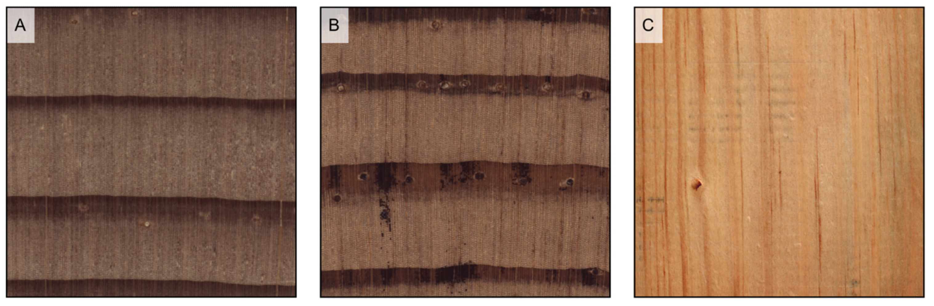
Earlywood/latewood transition. Pseudotsuga menziesii is an example of abrupt transition ((A1), transverse section) and evident wood figure is ((A2), tangential section); Cupressus sempervirens is an example of gradual transition ((B1), transverse section) and faint wood figure ((B2), tangential section).
Figure 5.
Earlywood/latewood transition. Pseudotsuga menziesii is an example of abrupt transition ((A1), transverse section) and evident wood figure is ((A2), tangential section); Cupressus sempervirens is an example of gradual transition ((B1), transverse section) and faint wood figure ((B2), tangential section).

Figure 6.
Wood porosity ((A–F) transverse sections, (G–I) tangential sections). (A) ring-porous, one row only of earlywood vessels, widest tangential spacing between earlywood vessels: more than one earlywood vessel (Carya ovata); (B) ring-porous, more than one row of earlywood vessels, widest tangential spacing between earlywood vessels: one earlywood vessel at most (Gleditsia triacanthos); (C) semi-ring porous, vessels’ diameter gradually narrows from earlywood to latewood (Juglans regia); (D) semi-ring porous, vessels are of the same diameter as latewood ones but much more closely spaced (Prunus avium); (E) diffuse porous, vessels are small (Acer rubrum); (F) diffuse porous, vessels are medium (Astronium graveolens). The figure consequent to growth rings is evident in ring-porous woods ((G) Fraxinus excelsior), visible in semi-ring porous woods ((H), Juglans regia), and faint in diffuse porous woods ((I), Betula pendula).

Figure 7.
Axial parenchyma distribution in softwoods (transverse sections). (A) scarce diffuse (Chamaecyparis lawsoniana); (B) tangentially zonate (Cryptomeria japonica); (C) absent (Araucaria angustifolia).
Figure 7.
Axial parenchyma distribution in softwoods (transverse sections). (A) scarce diffuse (Chamaecyparis lawsoniana); (B) tangentially zonate (Cryptomeria japonica); (C) absent (Araucaria angustifolia).

Figure 8.
Axial parenchyma distribution in hardwoods (transverse sections). (A) vasicentric (Khaya ivorensis); (B) lozenge-aliform, confluent, and in marginal or seemingly marginal bands (Afzelia bipindensis); (C) winged-aliform and confluent (Gonystylus bancanus); (D) diffuse (Alstonia macrophylla); (E) diffuse-in-aggregates and vasicentric (Dalbergia retusa); (F) narrow bands and reticulate (Autranella congolensis); (G) wide bands (Millettia laurentii); (H) festooned and scalariform (Grevillea robusta); (I) absent (Salix alba).

Figure 9.
If prominent enough, axial parenchyma appears on longitudinal surfaces as a light-colored band along the vessels’ grooves when paratracheal ((A) Afzelia bipindensis), and as seemingly growth rings when apotracheal in tangential bands ((B) Amphimas pterocarpoides).
Figure 9.
If prominent enough, axial parenchyma appears on longitudinal surfaces as a light-colored band along the vessels’ grooves when paratracheal ((A) Afzelia bipindensis), and as seemingly growth rings when apotracheal in tangential bands ((B) Amphimas pterocarpoides).

Figure 10.
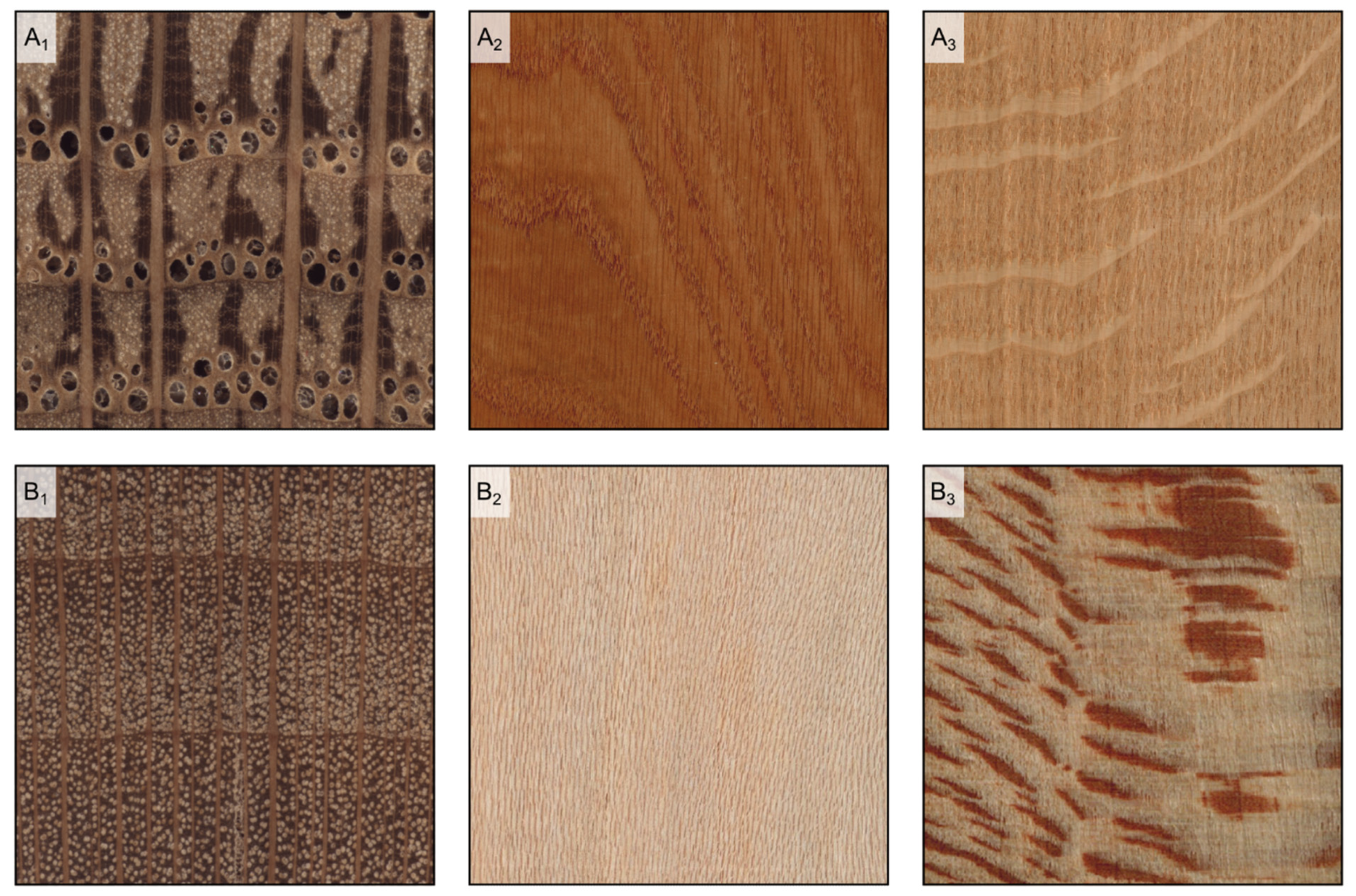
Rays’ visibility to the naked eye. In Quercus petraea, some of the rays are clearly more evident than the others on the transverse surface ((A1) transverse section), are visible on the tangential surface and over 5 mm high ((A2) longitudinal tangential surface), and visible on the radial surface ((A3) longitudinal radial surface); in Platanus occidentalis, rays are visible on the transverse surface and noded ((B1) transverse section), they are visible on the tangential surface and less than 5 mm high ((B2) longitudinal tangential surface), and visible on the radial surface ((B3) longitudinal radial surface); in Populus tremula, rays are not visible on either the transverse surface ((C1) transverse section) and the tangential surface ((C2) longitudinal tangential surface), while barely noticeable on the radial surface ((C3) longitudinal radial surface); in Pinus sylvestris, rays are not visible on either the transverse surface ((D1) transverse surface) and the tangential surface ((D2) tangential surface), while barely noticeable on the radial surface ((D3) longitudinal radial surface).
Figure 10.
Rays’ visibility to the naked eye. In Quercus petraea, some of the rays are clearly more evident than the others on the transverse surface ((A1) transverse section), are visible on the tangential surface and over 5 mm high ((A2) longitudinal tangential surface), and visible on the radial surface ((A3) longitudinal radial surface); in Platanus occidentalis, rays are visible on the transverse surface and noded ((B1) transverse section), they are visible on the tangential surface and less than 5 mm high ((B2) longitudinal tangential surface), and visible on the radial surface ((B3) longitudinal radial surface); in Populus tremula, rays are not visible on either the transverse surface ((C1) transverse section) and the tangential surface ((C2) longitudinal tangential surface), while barely noticeable on the radial surface ((C3) longitudinal radial surface); in Pinus sylvestris, rays are not visible on either the transverse surface ((D1) transverse surface) and the tangential surface ((D2) tangential surface), while barely noticeable on the radial surface ((D3) longitudinal radial surface).


Figure 11.
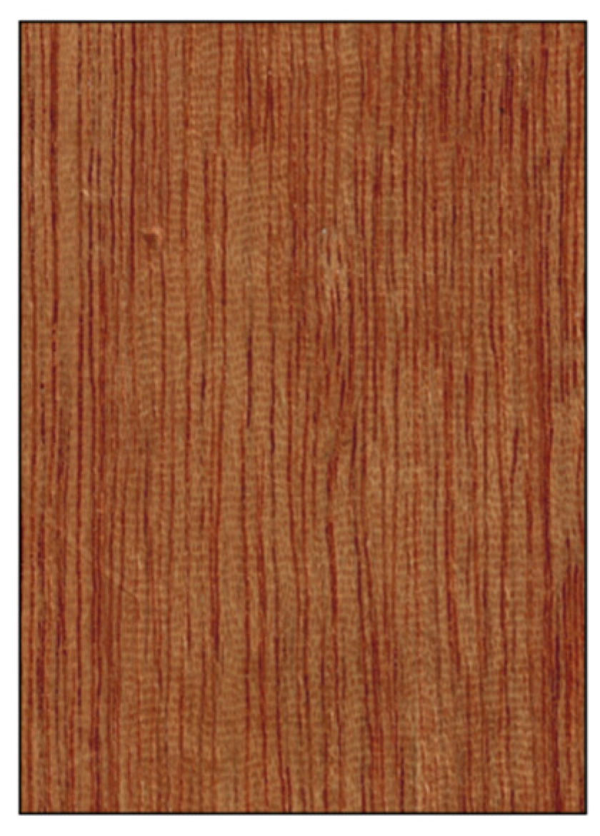
Ripple marks determined by storied rays on a tangential section of Swietenia macrophylla.
Figure 11.
Ripple marks determined by storied rays on a tangential section of Swietenia macrophylla.

Figure 12.
Vessels’ arrangement (all transverse sections except (E)). (A) tangential bands (latewood vessels only) (Ulmus glabra); (B) radial pattern (Ilex aquifolium); (C) diagonal pattern (Eucalyptus deglupta); (D) dendritic pattern (Bulnesia sarmientoi); (E) jagged pattern on longitudinal tangential surface determined by wavy tangential bands of latewood vessels (Ulmus glabra).

Figure 13.
Vessels’ groupings (transverse sections). (A) in radial multiples of 2–3 vessels (Hevea brasiliensis); (B) exclusively solitary (Quercus ilex); (C) radial multiples of 4 or more common (Dyera costulata); (D) clusters common (Celtis occidentalis).
Figure 13.
Vessels’ groupings (transverse sections). (A) in radial multiples of 2–3 vessels (Hevea brasiliensis); (B) exclusively solitary (Quercus ilex); (C) radial multiples of 4 or more common (Dyera costulata); (D) clusters common (Celtis occidentalis).

Figure 14.
Decorative grain deviations. (A) interlocked grain (Entandrophragma candollei); (B) wavy grain (Acer sp.); (C) quilt (Pterocaprus sp.); (D) burl (Pterocarpus indicus); (E) burl (Ulmus campestris); (F) crotch (Juglans regia).
Figure 14.
Decorative grain deviations. (A) interlocked grain (Entandrophragma candollei); (B) wavy grain (Acer sp.); (C) quilt (Pterocaprus sp.); (D) burl (Pterocarpus indicus); (E) burl (Ulmus campestris); (F) crotch (Juglans regia).

Figure 15.
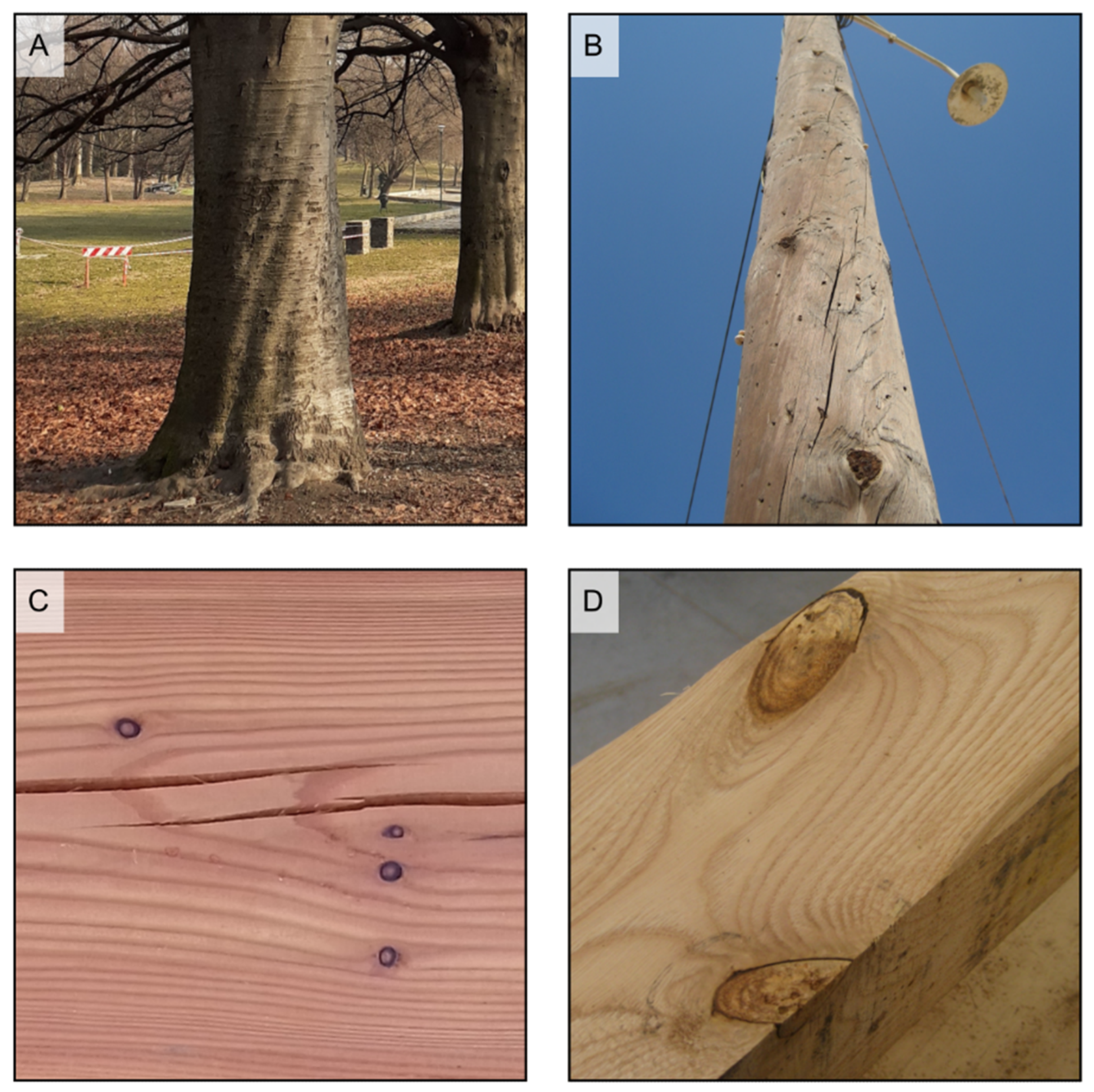
(A) spiral grain is visible in the tree in the foreground, while it is not present in that in the background (Fagus sp.); (B) spiral grain in a softwood pole. Grain deviates around knots ((C) small knots on Larix decidua; (D) pair of knots on Castanea sativa).
Figure 15.
(A) spiral grain is visible in the tree in the foreground, while it is not present in that in the background (Fagus sp.); (B) spiral grain in a softwood pole. Grain deviates around knots ((C) small knots on Larix decidua; (D) pair of knots on Castanea sativa).

Figure 16.
Vessel deposits (all transverse sections except (E), longitudinal section). Tyloses in the vessels of Robinia pseudoacacia (A) and Morus mesozygia (B); gums in the vessels of Swietenia humilis (C) and Lophira alata (D,E).
Figure 16.
Vessel deposits (all transverse sections except (E), longitudinal section). Tyloses in the vessels of Robinia pseudoacacia (A) and Morus mesozygia (B); gums in the vessels of Swietenia humilis (C) and Lophira alata (D,E).

Figure 17.
Axial canals in softwoods (all transverse sections except (C)). (A) axial canals small (Picea engelmannii); (B) axial canals large (Pinus rigida); (C) vertical streaks on a longitudinal surface determined by axial canals (Pinus chihuahuana).
Figure 17.
Axial canals in softwoods (all transverse sections except (C)). (A) axial canals small (Picea engelmannii); (B) axial canals large (Pinus rigida); (C) vertical streaks on a longitudinal surface determined by axial canals (Pinus chihuahuana).

Figure 18.
Axial canals in hardwoods (all transverse sections except (D)). (A) axial canals diffuse (Daniellia ogea); (B) axial canals in short tangential lines (Dipterocarpus alatus); (C) axial canals in long tangential lines (Shorea acuminata); (D) streaks on a longitudinal surface determined by axial canals (Prioria sp.).


Figure 19.
Traumatic canals as they appear on the transverse section of Scleronema micranthum (A) and Cedrus atlantica (B), and on the longitudinal section of Cariniana micrantha (C) and Eucalyptus obliqua (D).
Figure 19.
Traumatic canals as they appear on the transverse section of Scleronema micranthum (A) and Cedrus atlantica (B), and on the longitudinal section of Cariniana micrantha (C) and Eucalyptus obliqua (D).

Figure 20.
Diffuse included phloem on Aquilaria malaccensis ((A) transverse section) and pith flecks on Prunus serotina ((B) longitudinal section).
Figure 20.
Diffuse included phloem on Aquilaria malaccensis ((A) transverse section) and pith flecks on Prunus serotina ((B) longitudinal section).


Table 1.
List of macroscopic features for hardwood and softwood identification according to [4]. MFN = macroscopic feature number, P = present, A = absent, V = variable, NA = not applicable, TR = Transverse section, TLS = Tangential longitudinal section, RLS = Radial longitudinal section.
Table 1.
List of macroscopic features for hardwood and softwood identification according to [4]. MFN = macroscopic feature number, P = present, A = absent, V = variable, NA = not applicable, TR = Transverse section, TLS = Tangential longitudinal section, RLS = Radial longitudinal section.
| Structure | Property | MFN | Character | Character States | ||
|---|---|---|---|---|---|---|
| Anato-mical features | Hardwood | Growth rings | Growth rings | 1 | Growth rings distinct | P/A/V |
| 2 | Growth rings per cm | Numerical value/NA | ||||
| Vessels | Porosity | 3 | Diffuse porous | P/A/V | ||
| 4 | Semi-ring porous | P/A/V | ||||
| 5 | Ring-porous | P/A/V | ||||
| 6 | Number of rows of earlywood pores | One row/More than one row/V/NA | ||||
| 7 | Widest tangential spacing between earlywood vessels | One earlywood vessel at most/More than one earlywood vessel | ||||
| Arrangement | 8 | Vessels in tangential bands | P/A/V | |||
| 9 | Vessels in radial pattern | P/A/V | ||||
| 10 | Vessels in diagonal pattern (echelon) | P/A/V | ||||
| 11 | Vessels in dendritic pattern (flame-like) | P/A/V | ||||
| Groupings | 12 | Solitary and in radial multiples of 2–3 vessels | P/A/V | |||
| 13 | Exclusively solitary (90% or more) | P/A/V | ||||
| 14 | Radial multiples of 4 or more common | P/A/V | ||||
| 15 | Clusters common | P/A/V | ||||
| Frequency | 16 | ≤5 vessels per square mm | P/A/V | |||
| 17 | 6–20 vessels per square mm | P/A/V | ||||
| 18 | >20 vessels per square mm | P/A/V | ||||
| Vessel diameter/Pore visibility | 19 | Small (not visible to the naked eye, less than 80 μm) | P/A/V | |||
| 20 | Medium (just visible to the naked eye, 80–130 μm) | P/A/V | ||||
| 21 | Large (commonly visible to the naked eye, larger than 130 μm) | P/A/V | ||||
| Latewood pore visibility | 22 | Latewood pores large, individually distinct, and few enough that they can be readily counted | P/A/V/NA | |||
| Vesselless bands | 23 | Vesselless tangential bands | P/A | |||
| Tyloses | 24 | Tyloses common (TR, TLS, RLS) | P/A/V | |||
| Vessel deposits | 25 | Gums and other deposits in heartwood vessels (TR, TLS, RLS) | P/A/V | |||
| 26 | Deposits white (TR, TLS, RLS) | P/A | ||||
| 27 | Deposits yellow (TR, TLS, RLS) | P/A | ||||
| 28 | Deposits dark (TR, TLS, RLS) | P/A | ||||
| Axial parenchyma | Distribution | 29 | Diffuse | P/A/V | ||
| 30 | Diffuse-in-aggregates | P/A/V | ||||
| 31 | Vasicentric | P/A/V/Unilateral | ||||
| 32 | Lozenge-aliform | P/A/V/Unilateral | ||||
| 33 | Winged-aliform | P/A/V/Unilateral | ||||
| 34 | Confluent | P/A/V/Unilateral | ||||
| 35 | Banded | Majority wide/Majority narrow/V/A | ||||
| 36 | Banded parenchyma distribution | Throughout the ring/In latewood only/In earlywood only/NA | ||||
| 37 | Parenchyma bands wider than rays | P/A/V | ||||
| 38 | Parenchyma in marginal or seemingly marginal bands | P/A/V | ||||
| 39 | Reticulate | P/A/V | ||||
| 40 | Scalariform | P/A/V | ||||
| 41 | Festooned | P/A/V | ||||
| 42 | Predominant parenchyma pattern within the body of the ‘growth ring’ | A/Diffuse/Diffuse-in-aggregates/Vasicentric/Lozenge-aliform/Winged-aliform/Confluent/Ban-ded/In marginal or seemingly marginal bands/Reticulate/Scalariform/Festooned | ||||
| Rays | Width | 43 | Ray visibility to the naked eye on the transverse section | Rays not visible/Rays visible/Some of the rays clearly more evident than the others | ||
| 44 | Ray visibility with the naked eye on the tangential surface (TLS) | Rays not visible/Rays visible | ||||
| 45 | Ratio of ray width to pore diameter | Larger rays narrower than wider pores/Larger rays as wide or wider than wider pores | ||||
| 46 | Noded rays | P/A/V | ||||
| Storying | 47 | Ray storying (TLS) | Not storied (absent)/Regular coarse storying/Regular fine storying/Irregular coarse storying/Irregular fine storying | |||
| Height | 48 | Ray height (TLS) | Highest rays less than 5 mm high/Highest rays more than 5 mm high | |||
| Rays per mm | 49 | Rays per mm | ≤4 mm/5–12 mm/>12 mm/NA | |||
| Wood rayless | 50 | |||||
| Fibers | Arrangement | 51 | Fibers in radial arrangement | P/A | ||
| Canals | Intercellular canals | 52 | Axial canals | A/Diffuse/In short tangential lines/In long tangential lines/V | ||
| 53 | Traumatic canals | P | ||||
| 54 | Radial canals | P/A | ||||
| Phloem | Phloem | 55 | Included phloem | A/Diffuse/Concentric | ||
| Softwood | Growth rings | Earlywood/ Latewood transition | 56 | Earlywood/Latewood transition | Abrupt transition from earlywood to latewood/Gradual transition from earlywood to latewood/V | |
| Axial canals | Axial canals | 57 | Axial canals | Large/Small/A | ||
| 57b | Traumatic canals | P | ||||
| Axial parenchyma | Visibility | 58 | Axial parenchyma visible with hand lens | Scarce diffuse/Tangentially zonate/A | ||
| Non- anato-mical features | Hardwood + Softwood | Heartwood | Color | 59 | Heartwood color darker than sapwood color | P/A |
| 60 | Heartwood basically brown or shades of brown | P/A | ||||
| 61 | Heartwood basically red or shades of red | P/A | ||||
| 62 | Heartwood basically yellow or shades of yellow | P/A | ||||
| 63 | Heartwood basically white to gray | P/A | ||||
| 64 | Heartwood with streaks | P/A | ||||
| Density | 65 | Density | Density low: <0.40 g/cm3/Density medium: 0.40–0.75 g/cm3/Density high: >0.75 g/cm3 | |||
| Odor | 66 | Odor | A/Distinctly present and pleasant (sweet, spicy, floral)/Distinctly present and unpleasant (sour, bitter, fetid) | |||
| Oily surface | 67 | Oily surface | P/A | |||
| Habit | 68 | Tree | P/A/V | |||
| 69 | Shrub | P/A/V | ||||
| 70 | Vine/Liana/Climber | P/A/V | ||||
| Geographical distribution | 71 | Europe and temperate Asia (Brazier and Franklin region 74) | P/A | |||
| 72 | Europe, excluding Mediterranean | P/A | ||||
| 73 | Mediterranean including Northern Africa and Middle East | P/A | ||||
| 74 | Temperate Asia (China), Japan, USSR | P/A | ||||
| 75 | Central South Asia (Brazier and Franklin region 75) | P/A | ||||
| 76 | India, Pakistan, Sri Lanka | P/A | ||||
| 77 | Burma | P/A | ||||
| 78 | Southeast Asia and the Pacific (Brazier and Franklin region 76) | P/A | ||||
| 79 | Thailand, Laos, Vietnam, Cambodia (Indochina) | P/A | ||||
| 80 | Indomalesia: Indonesia, Philippines, Malaysia, Brunei, Singapore, Papua New Guinea, and Solomon Islands | P/A | ||||
| 81 | Pacific Islands (including New Caledonia, Samoa, Hawaii, and Fiji) | P/A | ||||
| 82 | Australia and New Zealand (Brazier and Franklin region 77) | P/A | ||||
| 83 | Australia | P/A | ||||
| 84 | New Zealand | P/A | ||||
| 85 | Tropical mainland Africa and adjacent islands (Brazier and Franklin region 78) | P/A | ||||
| 86 | Tropical Africa | P/A | ||||
| 87 | Madagascar & Mauritius, Reunion & Comores | P/A | ||||
| 88 | Southern Africa (south of the Tropic of Capricorn) (Brazier and Franklin region 79) | P/A | ||||
| 89 | North America, north of Mexico (Brazier and Franklin region 80) | P/A | ||||
| 90 | Neotropics and temperate Brazil (Brazier and Franklin region 81) | P/A | ||||
| 91 | Mexico and Central America | P/A | ||||
| 92 | Carribbean | P/A | ||||
| 93 | Tropical South America | P/A | ||||
| 94 | Southern Brazil | P/A | ||||
| 95 | Temperate South America including Argentina, Chile, Uruguay, and S. Paraguay (Brazier and Franklin region 82) | P/A | ||||
| Hardwood | Heartwood | Fluorescence | 96 | Surface fluorescence color | A/Basically yellow/Basically green/Other colors/V | |
| 97 | Surface fluorescence intensity | Weakly fluorescent/Strongly fluorescent/V/NA | ||||
| Extractives | 98 | Water extract fluorescence | A/Basically blue/Basically green/Bluish-green/V | |||
| 99 | Ethanol extract fluorescence | A/Basically blue/Basically green/Bluish-green/V | ||||
| 100 | Water extract color | Colorless/Brown or shades of brown/Red or shades of red/Yellow or shades of yellow/Other shades | ||||
| 101 | Ethanol extract color | Colorless/Brown or shades of brown/Red or shades of red/Yellow or shades of yellow/Other shades | ||||
| Froth test | 102 | Froth after shaking in water | Positive/Weakly positive/A | |||
| Burning splinter test | 103 | Splinter burns to: | Charcoal/Partial ash/Full ash (white)/Full ash (yellow-brown)/Full ash (other) | |||
| Chrome Azurol-S test | 104 | Chrome Azurol-S test | Positive/Negative | |||
| Grain | 105 | Interlocked grain | P | |||
| Surface marks | 106 | Surface marks | Pith flecks/Gum deposits/Kino veins/Pitch pockets/Latex traces/Resin veins | |||
Table 2.
List of recent references on macroscopic wood identification. The identification method is reported as traditional when performed by humans, and as machine vision when on an automated basis.
Table 2.
List of recent references on macroscopic wood identification. The identification method is reported as traditional when performed by humans, and as machine vision when on an automated basis.
| Name | Year | Area | Taxa (n) | Method (Traditional/Machine Vision) | References |
|---|---|---|---|---|---|
| Identification of Central American, Mexican, and Caribbean Woods | 2022 | Mexico, Central America, Caribbean | 138 species | Traditional | [16] |
| UTForest—UTFPR Classificador | 2021 | Brazil | 44 species | Traditional | [17] |
| Macroscopic wood identification key for Atlantic Forest species | 2021 | Brazil | 102 species | Traditional | [18] |
| Macroscopic wood identification key for Brazilian endangered species | 2021 | Brazil | 29 species | Traditional | [19] |
| Identification of Tree Species from the Peruvian Tropical Amazon “Selva Central” Forests According to Wood Anatomy | 2021 | Peru | 20 species | Traditional | [20] |
| Macroscopic wood identification key for Itatiaia National Park, RJ, Brazil | 2020 | Brazil | 41 species | Traditional | [21] |
| Field identification manual for Ghanaian timbers | 2020 | Ghana | 102 species | Traditional | [22] |
| Atlas of macroscopic wood identification: with a special focus on timbers used in Europe and CITES-listed species | 2019 | Global | 292 genera 335 species | Traditional | [3] |
| Forest Species Classifier | 2018 | Brazil | 112 species | Traditional | [23] |
| MacroHOLZdata | 2016 | Global | 150 species | Traditional | [24] |
| Forest Species Database—Macroscopic | 2014 | Brazil | 41 species | Traditional | [25] |
| Brazilian commercial timbers | 2010 | Brazil | 275 species | Traditional | [26] |
| CITESwoodID | 2005 | Various world regions | 75 species | Traditional | [13] |
| Towards Sustainable North American Wood Product Value Chains, Part I: Computer Vision Identification of Diffuse Porous Hardwoods | 2022 | North America | 24 genera 105 species | Machine vision | [27] |
| Towards sustainable North American wood product value chains, Part 2: computer vision identification of ring-porous hardwoods | 2022 | North America | 15 genera 68 species | Machine vision | [28] |
| Wood identification based on longitudinal section images by using deep learning | 2021 | North America | 11 species | Machine vision | [29] |
| Wood species automatic identification from wood core images with a residual convolutional neural network | 2021 | Europe | 14 species | Machine vision | [30] |
| Machine vision for field-level wood identification | 2020 | Amazonia Atlantic region | 21 species | Machine vision | [31] |
| Rapid field identification of CITES timber species by deep learning | 2020 | Suriname | 14 species | Machine vision | [32] |
| Software for forest species recognition based on digital images of wood | 2018 | Brazil | 41 species | Machine vision | [33] |
| Forest species recognition using deep convolutional neural networks | 2014 | Brazil | 41 species | Machine vision | [34] |
Table 3.
List of macroscopic features of softwoods and hardwoods.
| Macroscopic Feature | Softwoods | Hardwoods |
|---|---|---|
| Axial tracheids | Present | Rare (fibre tracheids s.l.) |
| Rays | Not visible to the naked eye on transverse and tangential planes | In several species visible to the naked eye on all three planes |
| Vessels | Absent | Present. In some species big enough to be seen by the naked eye |
| Fibers | Absent | Present |
| Canals | Present in some species | Present in some species |
| Appearance of growth rings | Distinct in most species | Variable |
Table 4.
Examples of species for different heartwood colors.
| Heartwood Color | Hardwoods | Softwoods |
|---|---|---|
| White | Acer spp., Carpinus betulus, Dyera costulata, Ilex aquifolium, Tilia cordata | Abies spp., Picea spp. |
| Brown | Carya ovata, Castanea sativa, Cordia dodecandra, Quercus spp., Tectona grandis | Araucaria spp., Pinus spp. |
| Yellow | Berberis vulgaris, Buxus sempervirens, Chloroxylon swietenia, Zanthoxylum flavum | Agathis spp., Cupressus spp. |
| Green | Cholorocardum rodiei, Guaiacum officinale, Liriodendron tulipifera, Handroanthus spp., Pistacia spp. | - |
| Red | Brosimum rubescens, Paubrasilia echinata, Pterocarpus soyauxii | Larix decidua, Taxus baccata |
| Orange | Centrolobium spp., Dalbergia retusa, Pterocarpus dalbergioides | - |
| Pink | Berchemya zeyheri, Dalbergia decipularis | - |
| Purple | Dalbergia cearensis, Peltogyne spp. | - |
| Black | Dalbergia melanoxylon, Diospyros crassiflora, Diospyros ebenum | - |
| Variegated | Astronium graveolens, Cordia dodecandra, Dalbergia nigra, Diospyros malabarica, Microberlinia brazzavillensis, Olea europaea, Zygia racemosa | - |
Table 5.
Examples of species for fine, medium, and coarse textures (these texture types are commonly reported in literature, for instance in [41]).
Table 5.
Examples of species for fine, medium, and coarse textures (these texture types are commonly reported in literature, for instance in [41]).
| Texture | Hardwood Species |
|---|---|
| Fine | Buxus sempervirens, Fagus sylvatica, Machaerium scleroxylon, Paubrasilia chinate, Pericospsis elata, Pyrus communis, Santalum album |
| Medium | Astromiun graveolens, Bowdichia nitida, Cordia dodecandra, Dipteryx odorata, Gonystylus bancanus, Mansonia altissima, Morus mesozygia |
| Coarse | Afzelia spp., Aucoumea klaineana, Castanea sativa, Dalbergia stevensonii, Hevea brasiliensis, Hymenaea courbaril, Terminalia superba |
Table 6.
Examples of species for different growth rings’ occurrence and visibility.
| Growth Rings | Hardwood Species |
|---|---|
| Distinct and clearly visible | Carya ovata, Cedrela odorata, Fagus sylvatica, Fraxinus excelsior, Juglans regia, Prunus avium, Quercus petraea, Tectona grandis, Ulmus glabra |
| Distinct but difficult to see | Eucalyptus camaldulensis, Malus sylvestris, Olea europaea, Platanus orientalis, Pyrus communis |
| Indistinct | Acacia koa, Brosimum alicastrum, Intsia bijuga, Khaya spp., Metopium brownei, Milicia spp., Millettia spp., Swietenia macrophylla |
| Variable | Aniba rosodora, Cordia dodecandra, Dalbergia cearensis, Dalbergia nigra, Santalum album, Tessmannia africana |
Table 7.
Examples of species for different types of transition in softwoods.
| Transition | Softwood Species |
|---|---|
| Abrupt | Larix decidua, Pinus spp. (hard pines group), Pseudotsuga menziesii, Tsuga heterophylla |
| Gradual | Abies alba, Agathis australis, Araucaria angustifolia, Calocedrus decurrens, Cedrus atlantica, Chamaecyparis lawsoniana, Cryptomeria japonica, Cupressus sempervirens, Pinus spp. (soft pines group), Taxus baccata |
| Variable | Fitzroya cupressoides, Picea abies, Sequoia sempervirens, Taxodium distichum, Thuja plicata |
Table 8.
Examples of species for different types of porosity in hardwoods.
| Porosity | Hardwood Species |
|---|---|
| Ring-porous | Carya ovata, Castanea sativa, Gleditsia triacanthos, Paulownia tomentosa, Quercus rubra, Robinia pseudoacacia, Ulmus minor |
| Semi-ring porous | Dalbergia decipularis, Juglans nigra, Juglans regia, Prunus avium, Prunus serotina, Pterocarpus indicus |
| Diffuse porous | Acer pseudoplatanus, Amphimas pterocarpoides, Betula spp., Handroanthus spp., Intsia bijuga, Liriodednron tulipifera, Magnolia ovata, Pyrus communis |
Table 9.
Examples of species for different types of axial parenchyma distribution in softwoods.
| Axial Parenchyma Distribution | Softwood Species |
|---|---|
| Scarce diffuse | Cedrus atlantica, Chamaecyparis lawsoniana |
| Tangentially zonate | Calocedrus decurrens, Cryptomeria japonica, Cupressus sempervirens, Fitzroya cupressoides, Podocarpus neriifolius, Thuja plicata |
| Absent | Abies alba, Araucaria angustifolia, Larix decidua, Picea abies, Pinus spp., Taxus baccata |
Table 10.
Examples of species for different types of axial parenchyma distribution in hardwoods.
| Axial Parenchyma Distribution | Hardwood Species |
|---|---|
| Absent | Acer pseudoplatanus, Alnus glutinosa, Buxus sempervirens, Casearia praecox, Euxylophora paraensis, Nothofagus pumilio, Populus spp. |
| Diffuse | Alstonia macrophylla, Aspidosperma polyneuron, Dillenia indica, Testulea gabonensis, Tetramerista glabra |
| Diffuse-in-aggregates | Carpinus betulus, Caryocar glabrum, Coula edulis, Dalbergia nigra, Heritiera utilis, Mammea Africana, Tilia cordata |
| Vasicentric | Acacia mangium, Antiaris toxicaria, Cordia dodecandra, Khaya ivorensis, Myroxylon balsamum, Newtonia leucocarpa |
| Lozenge-aliform | Afzelia spp., Berlinia bracteosa, Dipteryx odorata, Hymenaea courbaril, Koompassia malaccensis, Mangifera indica |
| Winged-aliform | Brosimum spp., Gonystylus spp., Jacaranda copaia, Poga oleosa, Simarouba amara, Tessmannia africana, Vochysia tetraphylla |
| Confluent | Andira coriacea, Dicorynia guianensis, Hymenolobium flavum, Leplaea cedrata, Milicia spp., Pterocaprus spp., Zygia racemosa |
| Narrow bands | Alstonia scholaris, Autranella congolensis, Cariniana legalis, Carya ovata, Diospyros spp., Dyera costulata, Hevea brasiliensis |
| Wide bands | Amphimas pterocarpoides, Clarisia racemosa, Ficus spp., Lophira alata, Millettia laurentii, Morus mesozygia, Swartzia cubensis |
| Marginal or seemingly marginal bands | Carapa guianensis, Chloroxylon swietenia, Gluta renghas, Swietenia macrophylla, Swintonia floribunda, Zanthoxylum flavum |
| Reticulate | Alstonia scholaris, Autranella congolensis, Cariniana legalis, Chrysophyllum africanum, Diospyros spp., Lophira alata |
| Scalariform | Ceiba pentandra, Dyera costulata, Grevillea robusta, Hexalobus crispiflorus, Roupala montana, Sterculia rhinopetala |
| Festooned | Grevillea robusta, Roupala montana |
Table 11.
Examples of species for different ray features in hardwoods.
| Ray Features | Hardwood Species |
|---|---|
| Rays not visible on the transverse surface | Autranella congolensis, Baikiaea plurijuga, Betula pendula, Dalbergia spp., Populus spp., Pterocarpus spp. |
| Rays visible on the transverse surface | Aucoumea klaineana, Celtis adolfi-friderici, Entandrophragma spp., Ilex aquifolium, Prunus avium |
| Some of the rays clearly more evident than the others on the transverse surface | Alnus glutinosa, Carpinus betulus, Fagus sylvatica, Gleditsia triacanthos, Quercus spp., Roupala montana |
| Rays not visible on the tangential surface | Afzelia spp., Dalbergia spp., Dyera costulata, Gluta renghas, Juglans regia, Salix spp. |
| Rays visible on the tangential surface, less than 5 mm high | Acer pseudoplatanus, Entandrophragma spp., Fagus sylvatica, Khaya spp., Platanus spp. |
| Rays visible on the tangential surface, over 5 mm high | Alnus glutinosa, Carpinus betulus, Quercus spp., Scaphium macropodum |
| Rays storied, fine | Dalbergia spp., Guaiacum officinale, Handroanthus spp., Machaerium scleroxylon, Pterocarpus spp. |
| Rays storied, coarse | Andira coriacea, Dipteryx odorata, Mansonia altissima, Millettia laurentii, Swietenia macrophylla |
Table 12.
Examples of species for different types of vessels’ arrangement.
| Vessels’ Arrangement | Hardwood Species |
|---|---|
| Tangential bands | Catalpa speciosa, Celtis australis, Gleditsia triacanthos, Grevillea robusta, Morus rubra, Roupala montana, Ulmus spp. |
| Radial pattern | Autranella congolensis, Baillonella toxisperma, Ilex aquifolium, Madhuca utilis, Manilkara bidentata, Palaquium obovatum |
| Diagonal pattern (echelon) | Bulnesia sarmientoi, Calophyllum canum, Eucalyptus spp., Mammea africana, Tieghemella heckelii |
| Dendritic pattern (flame-like) | Autranella congolensis, Bulnesia sarmientoi, Calophyllum canum, Castanea sativa, Quercus spp. |
Table 13.
Examples of species for different types of vessels’ groupings.
| Vessels’ Groupings | Hardwood Species |
|---|---|
| In radial multiples of 2–3 vessels | Acer pseudoplatanus, Amburana cearensis, Dalbergia spp., Erythrophleum ivorense, Fagus sylvatica, Hevea brasiliensis, Myroxylon balsamum |
| Exclusively solitary (90% or more) | Buxus sempervirens, Eucalyptus camaldulensis, Guaiacum officinale, Malus sylvestris, Quercus ilex, Santalum album |
| Radial multiples of 4 or more common | Alnus glutinosa, Aquilaria malaccensis, Baillonella toxisperma, Dyera costulata, Manilkara bidentata, Parahancornia fasciculata |
| Clusters common | Catalpa speciosa, Gleditsia triacanthos, Gymnocladus dioicus, Morus rubra, Robinia pseudoacacia, Ulmus spp. |
Table 14.
Examples of species for different vessel frequencies.
| Vessels’ Frequency | Hardwood Species |
|---|---|
| ≤5 vessels per square mm | Berlinia bracteosa, Dalbergia nigra, Hevea brasiliensis, Lophira alata, Ochroma pyramidale, Pterocarpus soyauxii |
| 6–20 vessels/square mm | Carapa guianensis, Entandrophragma cylindricum, Leplaea cedrata, Juglans regia, Swietenia macrophylla |
| >20 vessels/square mm | Acer spp., Betula spp., Dalbergia cearensis, Hopea ferrea, Liriodendron tulipifera, Prunus avium |
Table 15.
Examples of species that may present decorative figures due to grain deviations.
| Decorative Figure | Species |
|---|---|
| Interlocked | Chloroxylon swietenia, Entandrophragma cylindricum, Khaya spp., Lovoa trichilioides, Myroxylon balsamum, Turraeanthus africanus |
| Curly/Fiddleback | Acer spp., Chloroxylon swietenia, Entandrophragma cylindricum., Fraxinus spp., Guibourtia spp., Juglans regia |
| Quilt | Acer spp., Afzelia xylocarpa, Guibourtia tessmannii, Pterocarpus spp., Entandrophragma cylindricum. |
| Burl | Eucalyptus spp., Juglans regia, Pterocarpus indicus, Tetraclinis articulata, Ulmus spp. |
| Crotch | Amburana cearensis, Juglans spp., Swietenia spp. |
Table 16.
Examples of species for different types of vessel deposits.
| Vessel Deposits | Hardwood Species |
|---|---|
| Tyloses | Bagassa guianensis, Carya ovata, Eucalyptus spp., Gluta renghas, Morus mesozygia, Quercus petraea, Robinia psuedoacacia |
| Gums | Afzelia spp., Dalbergia spp., Intsia spp., Khaya spp., Lophira alata, Platymiscium spp., Pterocarpus spp., Swartzia spp. |
Table 17.
Examples of species for different types of axial canals’ distribution.
| Axial Canals’ Distribution | Hardwood Species |
|---|---|
| Diffuse | Anisoptera spp., Daniellia spp., Detarium spp., Dipterocarpus spp., Prioria spp., Tessmannia spp., Vatica spp. |
| In short tangential lines | Daniellia spp., Detarium spp., Dipterocarpus spp., Sindoropsis letestui, Tessmannia spp. |
| In long tangential lines | Copaifera spp., Eperua spp., Hopea spp., Neobalanocarpus heimii, Shorea spp., Sindora spp. |
Table 18.
Examples of species for different wood density classes.
| Wood Density | Species |
|---|---|
| <400 kg/m3 | Aquilaria malaccensis., Bombax spp., Calocedrus decurrens, Ceiba pentandra, Ochroma pyramidale, Paulownia tomentosa, Thuja plicata |
| 400–750 kg/m3 | Abies alba, Castanea sativa, Fagus sylvatica, Gmelina arborea, Juglans regia, Pinus spp., Quercus rubra, Tectona grandis |
| 760–1000 kg/m3 | Afzelia spp., Baikiaea plurijuga, Buxus sempervirens, Dalbergia nigra, Koompassia malaccensis, Olea europaea, Peltogyne spp. |
| >1000 kg/m3 | Brosimum guianense, Dalbergia melanoxylon, Guaiacum officinale, Handroanthus spp., Manilkara bidentata, Zollernia paraensis |
Table 19.
Examples of species for different wood odors.
| Wood Odor | Species |
|---|---|
| Present and pleasant (sweet, spicy, floral) | Aniba rosodora, Cedrela odorata, Cedrus spp., Cupressus spp., Juniperus spp., Myroxylon balsamum, Santalum album |
| Present and unpleasant (sour, bitter, fetid) | Alstonia congensis, Dinizia excelsa, Gonystylus spp., Microberlinia brazzavillensis, Piptadeniastrum africanum |
Table 20.
Examples of species with different heartwood fluorescence.
| Heartwood Fluorescence | Hardwood Species |
|---|---|
| Yellow, strong | Acacia mangium, Albizia ferruginea, Cedrelinga cateniformis, Dipteryx odorata, Robinia pseudoacacia |
| Yellow, weak | Brachystegia zenkeri, Guibourtia tessmannii, Mangifera indica, Pentaclethra macrophylla |
| Green, strong | Balfouridendron riedelianium, Dimorphandra polyandra |
| Green, weak | Centrolobium robustum, Dinizia excelsa |
Disclaimer/Publisher’s Note: The statements, opinions and data contained in all publications are solely those of the individual author(s) and contributor(s) and not of MDPI and/or the editor(s). MDPI and/or the editor(s) disclaim responsibility for any injury to people or property resulting from any ideas, methods, instructions or products referred to in the content. |
© 2023 by the authors. Licensee MDPI, Basel, Switzerland. This article is an open access article distributed under the terms and conditions of the Creative Commons Attribution (CC BY) license (https://creativecommons.org/licenses/by/4.0/).
Share and Cite
MDPI and ACS Style
Ruffinatto, F.; Negro, F.; Crivellaro, A. The Macroscopic Structure of Wood. Forests 2023, 14, 644. https://doi.org/10.3390/f14030644
AMA Style
Ruffinatto F, Negro F, Crivellaro A. The Macroscopic Structure of Wood. Forests. 2023; 14(3):644. https://doi.org/10.3390/f14030644
Chicago/Turabian StyleRuffinatto, Flavio, Francesco Negro, and Alan Crivellaro. 2023. "The Macroscopic Structure of Wood" Forests 14, no. 3: 644. https://doi.org/10.3390/f14030644
Note that from the first issue of 2016, this journal uses article numbers instead of page numbers. See further details here.