Article

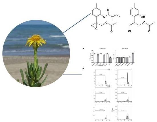
Bioassay-Guided Isolation of Antiproliferative Compounds from Limbarda crithmoides (L.) Dumort

by
Sabrina Adorisio
1,
Laura Giamperi
2,
Anahi Elena Ada Bucchini
2,
Domenico Vittorio Delfino
1,3,* and
Maria Carla Marcotullio
4,*
1
Department of Medicine, Foligno Nursing School, University of Perugia, 06034 Foligno, Italy
2
Department of Biomolecular Sciences, Section of Biochemistry and Biotechnology, University of Urbino, 61029 Urbino, Italy
3
Department of Medicine, Section of Pharmacology, University of Perugia, 06132 Perugia, Italy
4
Department of Pharmaceutical Sciences, Università degli Studi di Perugia, 06123 Perugia, Italy
*
Authors to whom correspondence should be addressed.
Molecules 2020, 25(8), 1893; https://doi.org/10.3390/molecules25081893
Submission received: 4 March 2020 / Revised: 16 April 2020 / Accepted: 17 April 2020 / Published: 20 April 2020
(This article belongs to the Special Issue Natural Products in Italy)

Abstract

:
Limbarda crithmoides (L.) Dumort (Asteraceae) n-hexane extract displayed high cell proliferation inhibitory activity against acute myeloid leukaemia cells (OCI-AML3) and was therefore subjected to a bioassay-guided multistep separation procedure. Two thymol derivatives, namely 10-acetoxy-8,9-epoxythymol tiglate (1) and 10-acetoxy-9-chloro-8,9-dehydrothymol (2), were isolated and identified by means of NMR spectroscopy. Both of them exhibited a significant dose-dependent inhibition of cell proliferation.

Graphical Abstract

1. Introduction

The genus Limbarda (Asteraceae), formerly included in the genus Inula, comprises two accepted species: L. crithmoides (L.) Dumort., and L. salsoloides (Turcz.) Ikonn [1]. L. crithmoides is a halophyte plant, commonly known as Inula crithmoides (L.), that is widespread across the salt marshes and sea cliffs of the Mediterranean Sea, French Atlantic coasts, English channel and western European seaboards [1]. L. crithmoides is a common edible plant. Its leaves, eaten raw or boiled, are a good source of protein, amino acids, fibers, vitamins and other components, and traditionally formed an important part of the Lebanese diet [2]. Young shoots of L. crithmoides are also consumed as pickles in vinegar in Spain [3], while its raw tops are added to salads in the Basilicata region of southern Italy [4]. I. crithmoides biological activities have been reviewed, along with other eight Asteraceae representative species [5]. Essential oil composition has been widely explored [6,7,8], and it showed to be dependent on the region where plants grow [8].
Extracts of L. crithmoides have been applied to crops and weeds to investigate their properties, which confirmed their herbicidal potency [9]. It was also reported that the extracts of callus cultures show good antifungal activity against Alternaria solani and Phytophthora cryptogea [10]. In a previous work, methanol and hexane extracts of the aerial parts of this plant were found to reduce the radial growth of A. solani and P. cryptogea, and they also show weak antifungal activity against fungi of the Fusarium genus [11]. The antioxidant activity of L. crithmoides has been widely investigated [11,12,13,14], and seems to be directly correlated with the presence of phenolic metabolites such as quinic acid derivatives [12,15,16,17]. For example, the ethyl acetate fraction of a methanolic extract of I. crithmoides showed in vitro and in vivo hepatoprotective activity against carbon tetrachloride (CCl4)-induced liver injury through antiradical and antioxidant activities [18]. In addition to antifungal activity, antimicrobial as well as antileishmanial activities of L. crithmoides extracts have also been reported [15,19,20,21].
As part of our ongoing research into antiproliferative compounds derived from natural sources [22,23,24,25,26], a phytochemical investigation of L. crithmoides was undertaken to isolate active compounds by means of a bioassay-guided fractionation of active extracts against the Ontario Cancer Institute-Acute Myeloid Leukemia-3 (OCI-AML3) acute myeloid leukaemia cell line.

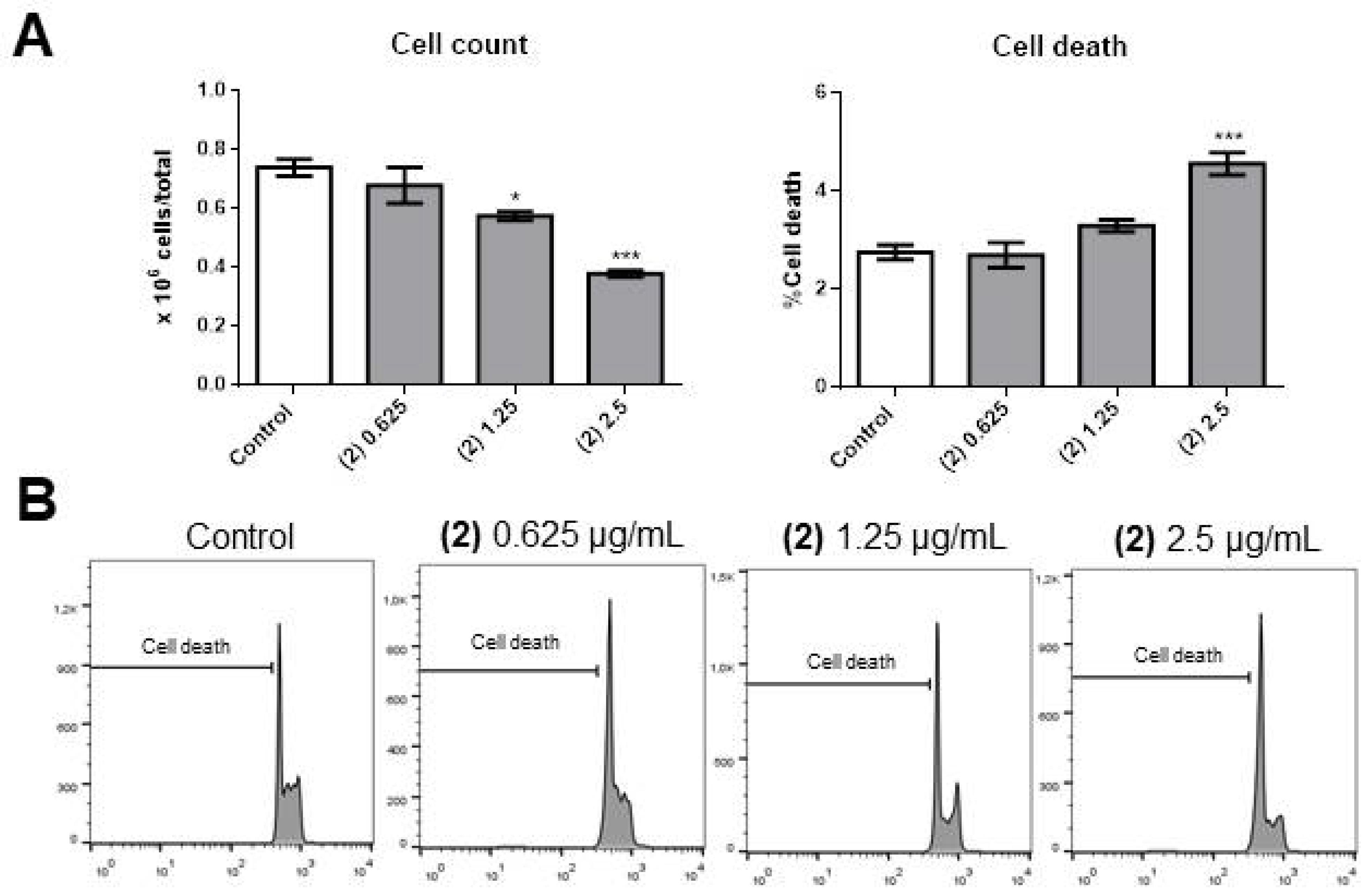
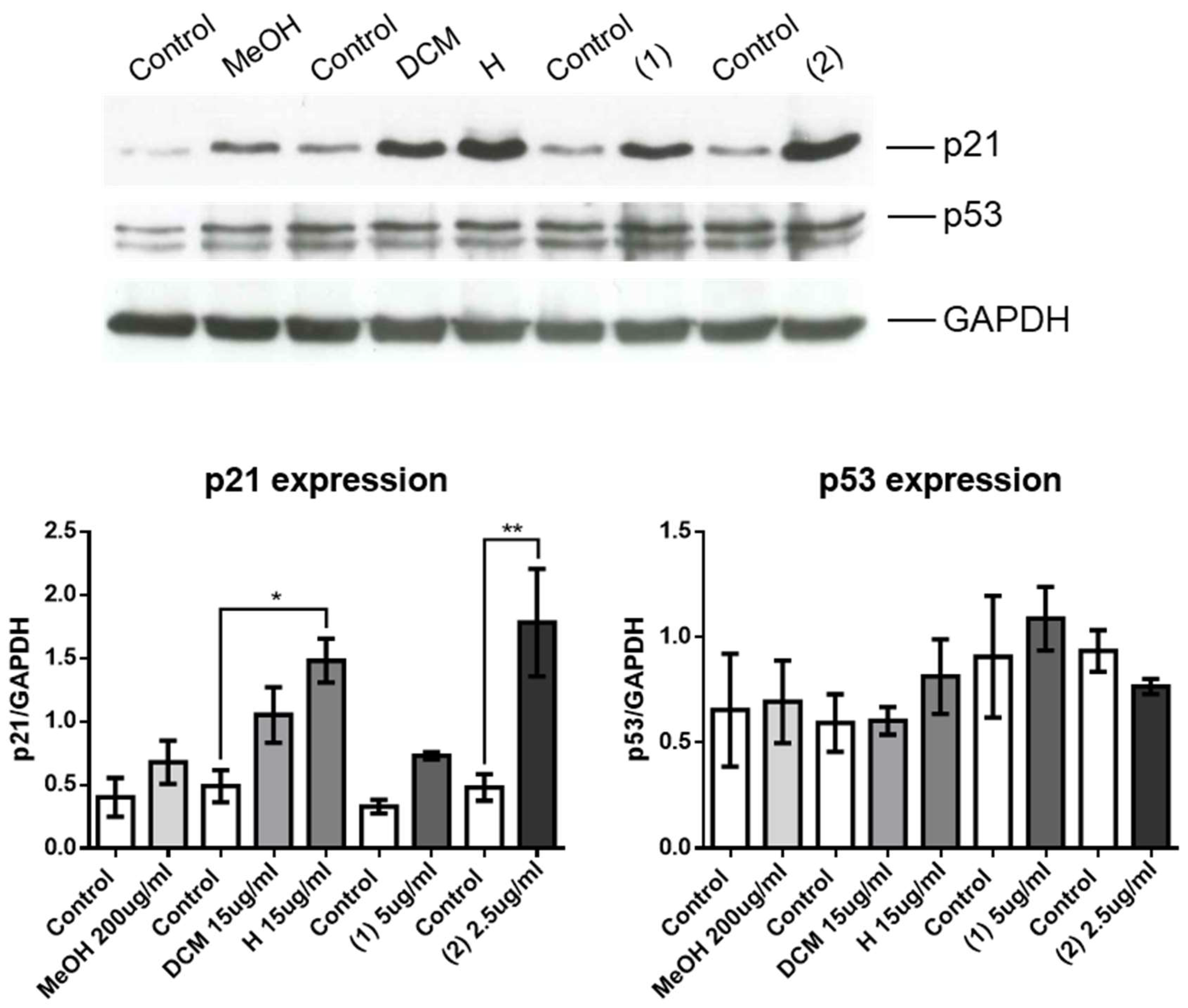
2. Results and Discussion

L. crithmoides (I. crithmoides) is a halophyte plant present in areas of high salinity [27,28] that represents an important agricultural crop [29,30] for its use as a food and for biological activities. Proximate composition of different extracts [6,7,31] showed the presence of saccharides [32] and proteins [33]. The most representative metabolites of this plant are phenolics [16,17,20,34,35,36,37] that are responsible of the good antioxidant and antiradical activity of extracts [10,11,12,13,15,38]. L. crithmoides essential oil and extracts were studied for their antimicrobial and antifungal activities [10,15,19,20,33,38]. Inula spp. have been investigated in depth for their cytotoxic/antiproliferative activity [39,40,41]. Despite the large number of biological studies undertaken on L. crithmoides [16,18,34,42], nothing has been reported so far about the cytotoxic/antiproliferative activity of this species and this prompted us to investigate the activity of L. crithmoides extracts against the OCI-AML3 cell line.
In a preliminary study, 24 h of treatment with 100 and 200 µg/mL of the methanolic extract (M) of L. crithmoides induced a significant decrease in OCI-AML3 cell number (Figure S1A, left panel, Supplementary Material). The augmentation observed with 200 µg/mL was accompanied by a significant increase in apoptosis (Figure S1A, right panel and S1B, Supplementary Material), associated with an accumulation of cells in the G0/G1 phase and a consequent decrease of cells in S and G2/M phases of the cell cycle (Figure S2A,B, Supplementary Material). The active M extract was subjected to solvent–solvent partitioning, yielding n-hexane (H), methylene chloride (DCM) and aqueous MeOH-soluble extracts. Both H and DCM extracts were able to significantly decrease the OCI-AML3 cell number compared to the vehicle (control) at concentrations of 15 or 10 μg/mL (with both extracts, Figure S3A, left panel, Supplementary Material). Analysis of apoptotic cell death showed a significant increase in apoptosis with 10 and 15 µg/mL of the H extract when compared with DCM extract (Figure S3A, right panel, and S3B, Supplementary Material). An analysis of the cell cycle revealed a significant increase in cells in the G0/G1 phase (Figure S4A, left panel, Supplementary Material) and fewer cells in S and G2/M phases of the cell cycle (Figure S4A, middle and right panels, respectively, Supplementary Material) with concentrations of 10 and 15 µg/mL for both fractions. Figure S4B shows a representative experiment of the experiment series quantified in Figure S4A. Thus, the H and DCM extracts were at least 10-fold more active than the M extract (Figures S3 and S4, Supplementary Material), and the H extract was more potent than the DCM extract in increasing apoptotic cell death. For this reason, we decided to continue only with the H extract.
The serial bioassay-guided chromatographic procedures led to the isolation of two active compounds (1 and 2; Figure 1). Using NMR spectroscopic data, compound 1 was identified as 10-acetoxy-8,9-epoxythymol tiglate, previously isolated from Athrixia spp. [43], Schkuhria multiflora Hook & Arn. [44] and Eupatorium cannabinum L. [45], and compound 2 was characterized as 10-acetoxy-9Z-chloro-8,9-dehydrothymol, previously isolated from Arnica sachalinensis (Regel) A. Gray [46] (Figures S5–S8, Supplementary Material). The Z configuration of the double bond was confirmed by NOESY experiment (Figure S9). Thymol derivatives are quite common in different Inula spp. [47,48,49], and it is known that L. crithmoides produces chlorinated thymol derivatives [37]. However, as far as we know, this is the first time that compounds 1 and 2 have been isolated from L. crithmoides (I. crithmoides). The two pure compounds were tested for their biological activity.
Compound 1 caused a significant decrease in OCI-AML3 cell number at 5 µg/mL (Figure 2A, left panel). This was possibly the consequence of a significant increase in apoptosis (Figure 2A, right panel, and Figure 2B) and blocking of cell cycle progression, as we observed significant accumulation of cells in the G0/G1 phase (Figure 3A, left panel) and fewer cells in the S (middle panel) and G2/M (right panel) phases of the cell cycle.
Compound 2 caused a significant decrease in cell number at concentrations as low as 1.25 µg/mL (Figure 4A, left panel), and there was a significant increase in apoptosis in cells treated with 2.5 µg/mL (Figure 4A, right panel, and Figure 4B). Compound 2 blocked the cell cycle progression at G0/G1 (Figure 5A, left panel), with 1.25 µg/mL causing a consequent significant decrease in cells in the S (Figure 5A, middle panel, and Figure 5B) and G2/M (Figure 5A, right panel, and Figure 5B) phases of the cell cycle.
Thus, compounds 1 and 2 were very effective in decreasing the number of OCI-AML3 cells through both increasing apoptosis and blocking cell proliferation. Compound 2 was more potent than compound 1 as it exerted inhibitory effects at a concentration as low as 1.25 µg/mL. The increased expression of p21 is largely associated with the arrest of cell cycle by two different pathways. The first is a p53-dependent and the second is p-53 -independent pathway. This prompted us to examine the expression of both p21 and p53 in OCI-AML3 cells untreated or treated with the M, DCM, H extracts and the compounds 1 and 2 isolated from the latter as a possible mechanism of cell cycle arrest since these pathways regulates mitotic progression and promotes cellular stress response [50]. We used Western blotting to measure expression of p21 in control and treated cells. As shown in Figure 6, upper panel, all the tested extracts and compounds induced an upregulation of p21, but only with the H extract and with compound 2 the increase of p21 was significant compared to controls (Figure 6, lower left panel). Because p21 is regulated through either p53-dependent or -independent pathways, we also measured expression of p53, which did not change after treatment with any of the extracts or compounds (Figure 6, upper and lower right panels).
These results suggest that the H extract induced a significant activation of a p21-dependent, p53-independent pathway in OCI-AML3 cells and that compound 2 was responsible of this activity and the associated cell cycle arrest. Notably, the p21-dependent, p-53-independent pathway has been shown to be associated with an increased apoptosis, as is evident in our system, in contrast to the p21-dependent, p53-dependent pathway that promotes the translocation of p21 from the nucleus to the cytoplasm thus determining an anti-apoptotic effect absent in our study [22].
It is known from previous studies that compounds isolated from Inula species may have antiproliferative effect on a different leukaemia cells [51,52,53,54,55,56,57,58,59] but, as far as we know, only one work describing the activity of Inula extract on an acute myeloid leukaemia (KG1a cell line) has been reported [60]. This work highlighted the effect of Inula compounds on induction of apoptosis by the mitochondria-dependent pathway. Most of the Inula metabolites tested were sesquiterpenolides. On the other hand, thymol and thymol derivatives as well as extracts and essential oils containing these compounds [61,62,63,64] are known to be cytotoxic.

3. Materials and Methods

3.1. General Chemical Material

NMR spectra were recorded using Avance DRX-400 and DPX-200 spectrometers (Bruker, Milan, Italy) operating at frequencies of 400 MHz (1H) and 100 MHz (13C) and 200 MHz (1H) and 50 MHz (13C), respectively The spectra were measured in CDCl3. The 1H- and 13C-NMR chemical shifts (δ) are expressed in ppm with reference to the solvent signals (CDCl3, δH 7.26 and δC 77.1). Coupling constants are given in Hz. NOESY (2D- NOE) experiments were executed on the Bruker Avance DRX-400 instrument. Preparative TLC was performed using pre-coated silica gel 60 F-254 plates (10 × 20 cm, Merck, Sigma-Aldrich, Milan, Italy) using n-hexane-acetone 8.5:1.5 as the eluent. Spots were visualized under UV light. Compounds were recovered from the stationary phase by washing five times with CH2Cl2 (DCM). Column chromatography was performed using MN Kiesegel 60 (70–230 mesh, Macherey-Nagel, Fisher Scientific, Milan, Italy). Fractions were monitored by TLC (Silica gel 60 F254; Merck), and spots on TLC were visualised under UV light and after staining with p-anisaldehyde-H2SO4-EtOH (1:1:98) followed by heating at 110 °C. All solvents used were of analytical grade and were purchased from VWR (VWR, Milan, Italy). Anhydrous Na2SO4 was purchased from Scharlau S.L. (Milan, Italy).

3.2. Plant Material

Aerial parts of L. crithmoides were collected during the flowering period (August 2016) in Fano, Urbino, Italy. Specimen collection was restricted to coastal habitats, nearly always within the reach of sea spray. Specimens were authenticated by Dr Laura Giamperi (University of Urbino). Voucher specimens have been deposited at the Herbarium of the Botanic Garden of the University of Urbino (GS 203).

3.3. Extraction and Isolation Procedure

Air-dried and finely powdered plant material (50 g) was extracted by maceration in methanol (MeOH) (3 × 500 mL for 24 h each). The combined extracts gave 12.87 g of active MeOH extract (M; yield 26%). The extract was dissolved in 10 mL of MeOH, diluted with H2O (100 mL), then subjected to solvent-solvent partition between n-hexane (3 × 50 mL; H extract; 6.2% yield) and methylene chloride (CH2Cl2) (3 × 50 mL; DCM extract; 3.4% yield). The H extract, which was the most active, was subjected to silica gel column chromatography (2 × 40 cm) under conditions of gradient elution using a mixture of CH2Cl2 in n-hexane (0→100%). Using n-hexane (1 L), we obtained four fractions: fraction 1 (IC-H-F1; 13 mg), using n-hexane-CH2Cl2 (1:1) (1.2 L) we obtained F2 (IC-H-F2, 280 mg), using CH2Cl2 (1.25 L) we obtained F3 (IC-H-F3, 120 mg) and F4 (IC-H-F4, 190 mg). Finally, the column was washed with 500 mL of MeOH (200 mg). The obtained fractions were tested for their antiproliferative activity using the same procedure for the extracts. An aliquot of the most active IC-H-F3 (109 mg) was further purified by silica gel column (1 × 20 cm) chromatography using CH2Cl2 as a solvent. Fractions (5 mL) were collected, evaluated by TLC and combined as a result of their similar appearance, yielding four pooled fractions: IC-H-F3(1), 36 mg; IC-H-F3(2), 52 mg; IC-H-F3(3), 6 mg; IC-H-F3(4), 6 mg. A solution of the IC-H-F3(1) fraction in 1 mL of DCM was purified by semipreparative TLC using n-hexane-acetone 8.5:1.5 as the eluent, resulting in the isolation of compound 1 (8 mg) and compound 2 (12 mg) (Figure S9, Supplementary Material). Each compound was identified by direct comparison of spectral properties, MS, 1H-NMR and 13C-NMR, with those of the authentic compounds in the literature [19,22].

3.4. Cell Line Culture and Characterisation

The antiproliferative activity of the two compounds under consideration was tested on the acute myeloid leukaemia (AML) cell line OCI-AML3 [65]. OCI-AML3 cells were maintained in Roswell Park Memorial Institute (RPMI) 1640 medium with 10% foetal bovine serum (FBS), 100 U/mL penicillin, and 100 μg/mL streptomycin at 37 °C and 5% CO2. Cells were purchased from ATCC (LGC Standards S.r.l., Sesto San Giovanni, Milan, Italy), kept at logarithmic growth and cultured in 24-well plates to assess their number and morphology. Cultures kept at 2 × 10⁵ cells/mL were treated with different concentrations of DMSO or the test compounds at the final concentrations reported in the figures. These reported concentrations were chosen based on preliminary experiments. After 24 h, the cell number was quantified using a haemocytometer.

3.5. Analysis of Cell Viability and Cell Cycle Progression

Cell viability and cell cycle progression were examined by flow cytometry to measure the amount of DNA in nuclei stained with propidium iodide (PI; Sigma-Aldrich, Milan, Italy), with the exclusion of necrotic cells by forward light scatter (FSC) [66]. Briefly, cells were harvested by centrifugation and gently resuspended in 1.5 mL hypotonic PI solution (50 µg/mL in 0.1% sodium citrate plus 0.1% Triton X-100). Tubes were kept in the dark at 4 °C for 30 min. PI fluorescence of individual nuclei was measured by flow cytometry using a Coulter Epics XL-MCLTM flow cytometer (Beckman Coulter, Cassina De’ Pecchi, Milan, Italy) and analysed using FlowJo_V10 software (BD Biosciences, Milan, Italy).

4. Conclusions

This study showed that the M extract of L. crithmoides has cell proliferation inhibitory activity against acute myeloid leukaemia cells (OCI-AML3) and solvent partition showed that the H fraction was more active than the DCM fraction. For this reason, we decided to continue testing the H fraction. Chromatographic purification of fraction H led to the isolation of two active thymol derivatives, 1 and 2. Compound 2 was shown to be highly active. As far as we know, this is the first report of the antiproliferative activity of chlorinated thymol derivatives. The DCM fraction, to a minor extent, was shown to still be active, and for this reason we think it would be interesting to explore its composition and the antiproliferative activity of its fractions and compounds.

Supplementary Materials

NMR spectra of the active compounds and biological activity assay result informations are available online.

Author Contributions

Conceptualization, M.C.M. and D.V.D.; plant material and preparation of extracts, L.G. and A.E.A.B.; chemical investigation, M.C.M.; biological investigation, S.A.; data curation, S.A.; writing—original draft preparation, M.C.M. and D.V.D.; writing—review and editing, M.C.M., D.V.D., L.G. and A.E.A.B.; supervision, M.C.M and D.V.D.; funding acquisition, D.V.D. All authors have read and agreed to the published version of the manuscript.

Funding

This research was funded by University of Perugia, Fondo Ricerca di Base 2019, Project “Studio sul processo di preparazione e validazione degli effetti biologici di sostanze naturali selezionate della medicina tradizionale Vietnamita”.

Acknowledgments

The manuscript has been revised by a professional language editing service (Proof-Reading-Service.com).

Conflicts of Interest

The authors declare no conflict of interest

References

  1. The Plant List: A Working List of All Plant Species. Available online: http://www.theplantlist.org (accessed on 10 October 2019).
  2. Zurayk, R.A.; Baalbaki, R. Inula crithmoides: A candidate plant for saline agriculture. Arid Soil Res. Rehabil. 1996, 10, 213–223. [Google Scholar] [CrossRef]
  3. Tardio, J.; Pardo de Santayana, M.; Morales, R. Ethnobotanical review of wild edible plants in Spain. Bot. J. Linn. Soc. 2006, 152, 27–71. [Google Scholar] [CrossRef]
  4. Guarrera, P.M.; Salerno, G.; Caneva, G. Food, flavouring and feed plant traditions in the Tyrrhenian sector of Basilicata, Italy. J. Ethnobiol. Ethnomed. 2006, 2, 37. [Google Scholar] [CrossRef] [PubMed] [Green Version]
  5. Bessada, S.M.F.; Barreira, J.C.M.; Oliveira, M.B.P.P. Asteraceae species with most prominent bioactivity and their potential applications: A review. Ind. Crop. Prod. 2015, 76, 604–615. [Google Scholar] [CrossRef]
  6. Harispe-Grandperrin, M.; Harispe, J.V. Proximate principles of Inula crithmoides L. family Compositae. II. Extraction and physical constants of the essential oil from the aerial parts. Bull. Soc. Chim. Biol. 1943, 25, 418–421. [Google Scholar]
  7. Harispe-Grandperrin, M.; Harispe, J.-V. Proximate principles of Inula crithmoides. III. Preliminary examination of the essential oil of the flower heads. Bull. Soc. Chim. Biol. 1944, 26, 192–202. [Google Scholar]
  8. Tsoukatou, M.; Roussis, V. Chemical composition and intra Mediterranean variation of the Inula crithmoides m L. oil. J. Essent. Oil Res. 1999, 11, 199–202. [Google Scholar] [CrossRef]
  9. Omezzine, F.; Daami-Remadi, M.; Rinez, A.; Ladhari, A.; Haouala, R. In vitro assessment of Inula spp. organic extracts for their antifungal activity against some pathogenic and antagonistic fungi. Afr. J. Microbiol. Res. 2011, 5, 3527–3531. [Google Scholar]
  10. Bucchini, A.; Giamperi, L.; Ricci, D. Total polyphenol content, in vitro antifungal and antioxidant activities of callus cultures from Inula crithmoides L. Nat. Prod. Commun. 2013, 8, 1587–1590. [Google Scholar] [CrossRef] [Green Version]
  11. Bucchini, A.; Ricci, D.; Messina, F.; Marcotullio, M.C.; Curini, M.; Giamperi, L. Antioxidant and antifungal activity of different extracts obtained from aerial parts of Inula crithmoides L. Nat. Prod. Res. 2015, 29, 1173–1176. [Google Scholar] [CrossRef]
  12. Jallali, I.; Waffo Teguo, P.; Smaoui, A.; Merillon, J.-M.; Abdelly, C.; Ksouri, R. Bio-guided fractionation and characterization of powerful antioxidant compounds from the halophyte Inula crithmoides. Arab. J. Chem. 2020, 13, 2680–2688. [Google Scholar] [CrossRef]
  13. Andreani, S.; De Cian, M.-C.; Paolini, J.; Desjobert, J.-M.; Costa, J.; Muselli, A. Chemical variability and antioxidant activity of Limbarda crithmoides L. essential oil from Corsica. Chem. Biodivers. 2013, 10, 2061–2077. [Google Scholar] [CrossRef] [PubMed]
  14. Giamperi, L.; Bucchini, A.; Fraternale, D.; Genovese, S.; Curini, M.; Ricci, D. Composition and antioxidant activity of Inula crithmoides essential oil grown in central Italy (Marche region). Nat. Prod. Commun. 2010, 5, 315–318. [Google Scholar] [CrossRef] [PubMed]
  15. Jallali, I.; Zaouali, Y.; Missaoui, I.; Smeoui, A.; Abdelly, C.; Ksouri, R. Variability of antioxidant and antibacterial effects of essential oils and acetonic extracts of two edible halophytes: Crithmum maritimum L. and Inula crithmoides L. Food Chem. 2014, 145, 1031–1038. [Google Scholar] [CrossRef]
  16. Aboul Ela, M.A.; El-Lakany, A.M.; Abdel-Kader, M.S.; Alqasoumi, S.I.; Shams-El-Din, S.M.; Hammoda, H.M. New quinic acid derivatives from hepatoprotective Inula crithmoides root extract. Helv. Chim. Acta 2012, 95, 61–66. [Google Scholar]
  17. Males, Z.; Plazibat, M.; Greiner, M. Qualitative and quantitative analysis of flavonoids of golden samphire -Limbarda crithmoides (L.) Dumort. Farm. Glas. 2004, 60, 453–459. [Google Scholar]
  18. Malash, B.N.; Ibrahim, S.M.; Ibrahim, A.-R.S.; Kabbash, A.; El-Aasr, M. In vitro and in vivo hepatoprotective study of Inula crithmoides L. Pluchea dioscoridis (L.) Desf. and Phyllanthus reticulates Poir. J. Pharm. Sci. Res. 2015, 7, 987–993. [Google Scholar]
  19. Assi, M.; Aboul-Ela, M.A.; Ellakany, A.M.; Abdul-Ghani, M. A comparative phytochemical and antimicrobial analysis of Inula viscosa and Inula crithmoides grown in Lebanon. Acad. J. Med. Plants 2014, 2, 057–067, 11 pp. [Google Scholar]
  20. Aboul Ela, M.A.; El-Lakany, A.M.; Shams-El-Din, S.M.; Hammoda, H.M. Phytochemical and antimicrobial investigation of Inula crithmoides L. Alexandria J. Pharm. Sci. 2011, 25, 37–40. [Google Scholar]
  21. Oliveira, M.; Joao Rodrigues, M.; Pereira, C.; Neto, R.L.D.M.; Junior, P.A.S.; Neng, N.D.R.; Nogueira, J.M.F.; Varela, J.; Barreira, L.; Custodio, L. First report of the in vitro antileishmanial properties of extremophile plants from the Algarve Coast. Nat. Prod. Res. 2018, 32, 600–604. [Google Scholar] [CrossRef]
  22. Adorisio, S.; Fierabracci, A.; Gigliarelli, G.; Muscari, I.; Cannarile, L.; Liberati, A.M.; Marcotullio, M.C.; Riccardi, C.; Curini, M.; Robles Zepeda, R.E.; et al. The hexane fraction of Bursera microphylla A Gray induces p21-mediated antiproliferative and proapoptotic effects in human cancer-derived cell lines. Integr. Cancer Ther. 2017, 16, 426–435. [Google Scholar] [CrossRef] [PubMed] [Green Version]
  23. Gigliarelli, G.; Zadra, C.; Cossignani, L.; Robles Zepeda, R.E.; Rascon-Valenzuela, L.A.; Velazquez-Contreras, C.A.; Marcotullio, M.C. Two new lignans from the resin of Bursera microphylla A. Gray and their cytotoxic activity. Nat. Prod. Res. 2018, 32, 2646–2651. [Google Scholar] [CrossRef] [PubMed]
  24. Marcotullio, M.C.; Loizzo, M.R.; Messina, F.; Temperini, A.; Tundis, R.; Menichini, F.; Curini, M. Bioassay-guided fractionation of Euphrasia pectinata Ten. and isolation of iridoids with antiproliferative activity. Phytochem. Lett. 2015, 12, 252–256. [Google Scholar] [CrossRef]
  25. Messina, F.; Curini, M.; Di Sano, C.; Zadra, C.; Gigliarelli, G.; Rascon-Valenzuela, L.A.; Robles Zepeda, R.E.; Marcotullio, M.C. Diterpenoids and triterpenoids from the Resin of Bursera microphylla and their cytotoxic activity. J. Nat. Prod. 2015, 78, 1184–1188. [Google Scholar] [CrossRef] [PubMed]
  26. Torres-Moreno, H.; Velazquez, C.A.; Garibay-Escobar, A.; Curini, M.; Marcotullio, M.C.; Robles-Zepeda, R.E. Antiproliferative and apoptosis induction of cucurbitacin-type triterpenes from Ibervillea sonorae. Ind. Crop. Prod. 2015, 77, 895–900. [Google Scholar] [CrossRef]
  27. Al Hassan, M.; Chaura, J.; López-Gresa, M.P.; Borsai, O.; Daniso, E.; Donat-Torres, M.P.; Mayoral, O.; Vicente, O.; Boscaiu, M. Native-Invasive Plants vs. Halophytes in Mediterranean Salt Marshes: Stress Tolerance Mechanisms in Two Related Species. Front. Plant Sci. 2016, 7, 473. [Google Scholar] [CrossRef] [Green Version]
  28. Gil, R.; Bautista, I.; Boscaiu, M.; Lidón, A.; Wankhade, S.; Sánchez, H.; Llinares, J.; Vicente, O. Responses of five Mediterranean halophytes to seasonal changes in environmental conditions. AoB Plants 2014, 6, plu049. [Google Scholar] [CrossRef] [PubMed]
  29. Ventura, Y.; Eshel, A.; Pasternak, D.; Sagi, M. The development of halophyte-based agriculture: Past and present. Ann. Bot. 2015, 115, 529–540. [Google Scholar] [CrossRef] [Green Version]
  30. Fita, A.; Rodríguez-Burruezo, A.; Boscaiu, M.; Prohens, J.; Vicente, O. Breeding and Domesticating Crops Adapted to Drought and Salinity: A New Paradigm for Increasing Food Production. Front. Plant Sci. 2015, 6, 978. [Google Scholar] [CrossRef] [Green Version]
  31. Harispe-Grandperrin, M.; Harispe, J.V. Proximate principles of Inula crithmoides L. family Compositae. I. Biochemical study of glucide constituents. Bull. Soc. Chim. Biol. 1943, 25, 416–418. [Google Scholar]
  32. Kacem, I.; Majdoub, H.; Roudesli, S. Fraction of soluble polysaccharides from Inula crithmoides by sequential extraction. J. Appl. Sci. 2008, 8, 2442–2448. [Google Scholar] [CrossRef]
  33. Attard, E.; Pacioni, P. The Phytochemical and In Vitro Pharmacological Testing of Maltese Medicinal Plants. In Bioactive Compounds in Phytomedicine; InTech: London, UK, 2011; pp. 93–112. [Google Scholar]
  34. Selim, M.A.; El-Hefnawy, H.M.; Ibrahim, T.A.; Sleem, A.A. Flavonoids and antihepatotoxic activity of Inula crithmoides L. growing in Egypt. Egypt. J. Biomed. Sci. 2003, 12, 83–95. [Google Scholar]
  35. El-Lakany, A.M.; Aboul Ela, M.A.; Hammoda, H.M.; Ghazy, N.M.; Mahmoud, Z.F. New methoxylated flavonols from Inula crithmoides. Pharmazie 1996, 51, 435–436. [Google Scholar]
  36. Metwally, M.A.; Dawidar, A.M. A thymol derivative from Inula crithmoides. Phytochemistry 1985, 24, 1377–1378. [Google Scholar] [CrossRef]
  37. Marco, J.A.; Sanz-Cervera, J.F.; Manglano, E. Chlorinated thymol derivatives from Inula crithmoides. Phytochemistry 1993, 33, 875–878. [Google Scholar] [CrossRef]
  38. Jdey, A.; Falleh, H.; Ben Jannet, S.; Mkadmini Hammi, K.; Dauvergne, X.; Ksouri, R.; Magne, C. Phytochemical investigation and antioxidant, antibacterial and anti-tyrosinase performances of six medicinal halophytes. S. Afr. J. Bot. 2017, 112, 508–514. [Google Scholar] [CrossRef]
  39. Seca, A.M.L.; Grigore, A.; Pinto, D.C.G.A.; Silva, A.M.S. The genus Inula and their metabolites: From ethnopharmacological to medicinal uses. J. Ethnopharmacol. 2014, 154, 286–310. [Google Scholar] [CrossRef] [Green Version]
  40. Wang, G.-W.; Qin, J.-J.; Cheng, X.-R.; Shen, Y.-H.; Shan, L.; Jin, H.-Z.; Zhang, W.-D. Inula sesquiterpenoids: Structural diversity, cytotoxicity and anti-tumor activity. Expert Opin. Investig. Drugs 2014, 23, 317–345. [Google Scholar] [CrossRef] [PubMed]
  41. Trendafilova, A.; Ivanova, V.; Rangelov, M.; Todorova, M.; Ozek, G.; Yur, S.; Ozek, T.; Aneva, I.; Veleva, R.; Moskova-Doumanova, V.; et al. Caffeoylquinic Acids, Cytotoxic, Antioxidant, Acetylcholinesterase and Tyrosinase Enzyme Inhibitory Activities of Six Inula Species from Bulgaria. Chem. Biodivers. 2020. Ahead of Print. [Google Scholar] [CrossRef]
  42. Abdel-Wahhab, M.A.; Abdel-Azim, S.H.; El-Nekeety, A.A. Inula crithmoides extract protects against ochratoxin A-induced oxidative stress, clastogenic and mutagenic alterations in male rats. Toxicon 2008, 52, 566–573. [Google Scholar] [CrossRef]
  43. Bohlmann, F.; Zdero, C. Neue norkauren- und thymol-derivate aus Athrixia-arten. Phytochemistry 1977, 16, 1773–1776. [Google Scholar] [CrossRef]
  44. Pacciaroni, A.D.V.; Sosa, V.E.; Espinar, L.A.; Oberti, J.C. Acyl glucopyranosides and other constituents from Schkuhria multiflora. An. Asoc. Quim. Argent. 1998, 86, 256–260. [Google Scholar]
  45. Chen, J.-J.; Tsai, Y.-C.; Hwang, T.-L.; Wang, T.-C. Thymol, Benzofuranoid, and Phenylpropanoid Derivatives: Anti-inflammatory Constituents from Eupatorium cannabinum. J. Nat. Prod. 2011, 74, 1021–1027. [Google Scholar] [CrossRef] [PubMed]
  46. Passreiter, C.M.; Matthiesen, U.; Willuhn, G. 10-Acetoxy-9-chloro-8,9-dehydrothymol and further thymol derivatives from Arnica sachalinensis. Phytochemistry 1998, 49, 777–778. [Google Scholar] [CrossRef]
  47. Wang, C.; Zhang, X.; Wei, P.; Cheng, X.; Ren, J.; Yan, S.; Zhang, W.; Jin, H. Chemical constituents from Inula wissmanniana and their anti-inflammatory activities. Arch. Pharm. Res. 2013, 36, 1516–1524. [Google Scholar] [CrossRef] [PubMed]
  48. Cheng, X.-R.; Ye, J.; Ren, J.; Zeng, Q.; Zhang, F.; Qin, J.-J.; Shen, Y.-H.; Zhang, W.-D.; Jin, H.-Z. Terpenoids from Inula sericophylla Franch. and their chemotaxonomic significance. Biochem. Syst. Ecol. 2012, 42, 75–78. [Google Scholar] [CrossRef]
  49. Zhao, J.; Li, Y.; Liu, Q.; Gao, K. Antimicrobial activities of some thymol derivatives from the roots of Inula hupehensis. Food Chem. 2010, 120, 512–516. [Google Scholar] [CrossRef]
  50. Adorisio, S.; Fierabracci, A.; Muscari, I.; Liberati, A.M.; Cannarile, L.; Thuy, T.T.; Sung, T.V.; Sohrab, H.; Hasan, C.M.; Ayroldi, E.; et al. Fusarubin and Anhydrofusarubin Isolated from A Cladosporium Species Inhibit Cell Growth in Human Cancer Cell Lines. Toxins 2019, 11, 503. [Google Scholar] [CrossRef] [Green Version]
  51. Yami, A.; Hamzeloo-Moghadam, M.; Darbandi, A.; Karami, A.; Mashati, P.; Takhviji, V.; Gharehbaghian, A. Ergolide, a potent sesquiterpene lactone induces cell cycle arrest along with ROS-dependent apoptosis and potentiates vincristine cytotoxicity in ALL cell lines. J. Ethnopharmacol. 2020, 253, 112504. [Google Scholar] [CrossRef]
  52. Karami, A.; Hamzeloo-Moghadam, M.; Yami, A.; Barzegar, M.; Mashati, P.; Gharehbaghian, A. Antiproliferative Effect of Gaillardin from Inula oculus-christi in Human Leukemic Cells. Nutr. Cancer 2019, 1–14. [Google Scholar] [CrossRef]
  53. Xu, X.; Huang, L.; Zhang, Z.; Tong, J.; Mi, J.; Wu, Y.; Zhang, C.; Yan, H. Targeting non-oncogene ROS pathway by alantolactone in B cell acute lymphoblastic leukemia cells. Life Sci. 2019, 227, 153–165. [Google Scholar] [CrossRef] [PubMed]
  54. Cai, H.; Li, L.; Jiang, J.; Zhao, C.; Yang, C. Costunolide enhances sensitivity of K562/ADR chronic myeloid leukemia cells to doxorubicin through PI3K/Akt pathway. Phytother. Res. 2019, 33, 1683–1688. [Google Scholar] [CrossRef] [PubMed]
  55. Cai, H.; Meng, X.; Li, Y.; Yang, C.; Liu, Y. Growth inhibition effects of isoalantolactone on K562/A02 cells: Caspase-dependent apoptotic pathways, S phase arrest, and downregulation of Bcr/Abl. Phytother. Res. 2014, 28, 1679–1686. [Google Scholar] [CrossRef] [PubMed]
  56. Pal, H.C.; Sehar, I.; Bhushan, S.; Gupta, B.D.; Saxena, A.K. Activation of caspases and poly (ADP-ribose) polymerase cleavage to induce apoptosis in leukemia HL-60 cells by Inula racemosa. Toxicol. Vitr. 2010, 24, 1599–1609. [Google Scholar] [CrossRef] [PubMed]
  57. Zeng, G.Z.; Tan, N.H.; Ji, C.J.; Fan, J.T.; Huang, H.Q.; Han, H.J.; Zhou, G.B. Apoptosis inducement of bigelovin from Inula helianthus-aquatica on human Leukemia U937 cells. Phytother. Res. 2009, 23, 885–891. [Google Scholar] [CrossRef]
  58. Pan, M.H.; Chiou, Y.S.; Cheng, A.C.; Bai, N.; Lo, C.Y.; Tan, D.; Ho, C.T. Involvement of MAPK, Bcl-2 family, cytochrome c, and caspases in induction of apoptosis by 1,6-O,O-diacetylbritannilactone in human leukemia cells. Mol. Nutr. Food Res. 2007, 51, 229–238. [Google Scholar] [CrossRef]
  59. Yue, G.G.; Chan, B.C.; Kwok, H.F.; Wong, Y.L.; Leung, H.W.; Ji, C.J.; Fung, K.P.; Leung, P.C.; Tan, N.H.; Lau, C.B. Anti-angiogenesis and immunomodulatory activities of an anti-tumor sesquiterpene bigelovin isolated from Inula helianthus-aquatica. Eur. J. Med. Chem. 2013, 59, 243–252. [Google Scholar] [CrossRef]
  60. Ding, Y.; Pan, W.; Xu, J.; Wang, T.; Chen, T.; Liu, Z.; Xie, C.; Zhang, Q. Sesquiterpenoids from the roots of Inula helenium inhibit acute myelogenous leukemia progenitor cells. Bioorg. Chem. 2019, 86, 363–367. [Google Scholar] [CrossRef]
  61. Elbe, H.; Yigitturk, G.; Cavusoglu, T.; Uyanikgil, Y.; Ozturk, F. Apoptotic effects of thymol, a novel monoterpene phenol, on different types of cancer. Bratisl. Lek. Listy 2020, 121, 122–128. [Google Scholar] [CrossRef] [Green Version]
  62. Yu, Y.; Liu, Y.; Shi, R.; Zhang, D.; Li, C.; Shi, J. New thymol and isothymol derivatives from Eupatorium fortunei and their cytotoxic effects. Bioorg. Chem. 2020, 103644. [Google Scholar] [CrossRef]
  63. De La Chapa, J.J.; Singha, P.K.; Lee, D.R.; Gonzales, C.B. Thymol inhibits oral squamous cell carcinoma growth via mitochondria-mediated apoptosis. J. Oral Pathol. Med. 2018, 47, 674–682. [Google Scholar] [CrossRef] [PubMed]
  64. Gunes-Bayir, A.; Kocyigit, A.; Guler, E.M. In vitro effects of two major phenolic compounds from the family Lamiaceae plants on the human gastric carcinoma cells. Toxicol. Ind. Health 2018, 34, 525–539. [Google Scholar] [CrossRef] [PubMed]
  65. Thuy, T.T.; Huong, N.T.T.; Nhung, L.T.H.; Ninh, P.T.; Delfino, D.V.; Van Sung, T. Isolation, characterization and biological evaluation of a phenoxazine, a natural dyestuff isolated from leaves of Peristrophe bivalvis. Nat. Prod. Res. 2013, 27, 771–774. [Google Scholar] [CrossRef]
  66. Riccardi, C.; Nicoletti, I. Analysis of apoptosis by propidium iodide staining and flow cytometry. Nat. Protoc. 2006, 1, 1458–1461. [Google Scholar] [CrossRef] [PubMed]
Sample Availability: Samples of the compounds are not available from the authors.
Figure 1. Chemical structures of active compounds 1 and 2.
Molecules 25 01893 g001
Figure 2. Effects of compound 1 on OCI-AML3 cell number and apoptotic cell death. (A) Bars represent the cell number (left panel) or the percentage of apoptotic cells after 24 h of treatment with control vehicle (Control) or 1,25 [(1) 1.25)], 2,5 [(1) 2.5] or 5 [(1) 5] µg/mL of compound 1. (B) Flow cytometry analyses of a representative experiment. Data from three independent experiments are reported as mean ± SEM. *** p < 0.001.
Molecules 25 01893 g002
Figure 3. Effects of compound 1 on OCI-AML3 cell cycle progression. (A) Bars represent the percentage of cells in G0/G1 (left panel), S (middle panel), or G2/M (right panel) phases after 24 h of treatment with control vehicle (Control) or 1.25 [(1) 1.25], 2.5 [(1) 2.5] or 5 [(1) 5] µg/mL of compound 1. (B) Flow cytometry analyses of a representative experiment. Data from three independent experiments are reported as mean ± SEM. * p < 0.05, ** p < 0.01.
Molecules 25 01893 g003
Figure 4. Effects of compound 2 on OCI-AML3 cell number and apoptotic cell death. (A) Bars represent the cell number (left panel) or the percentage of apoptotic cells after 24 h of treatment with control vehicle (Control) or 0.625 [(2) 0.625)], 1.25 [(2) 1.25] or 2.5 [(2) 2.5] µg/mL of compound 2. (B) Flow cytometry analyses of a representative experiment. Data from three independent experiments are reported as mean ± SEM. * p < 0.05; *** p < 0.001.
Molecules 25 01893 g004
Figure 5. Effects of compound 2 on OCI-AML3 cell cycle progression. (A) Bars represent the percentage of cells in G0/G1 (left panel), S (middle panel), or G2/M (right panel) phases after 24 h of treatment with control vehicle (Control) or 0.625 [(2) 0.625], 1.25 [(2) 2.25] or 2.5 [(2) 2.5] µg/mL of compound 2. (B) Flow cytometry analyses of a representative experiment. Data from three independent experiments are reported as mean ± SEM. * p < 0.05, ** p < 0.01.
Molecules 25 01893 g005aMolecules 25 01893 g005b
Figure 6. Effects of extracts and compounds on expression of proteins involved in the cell cycle. Upper panel, western blot analysis illustrating expression of p21, p53 and GAPDH using cell lysates extracted from OCI-AML3 cells treated with vehicle (Control), M (MeOH), DCM (DCM), H (H) extracts and compound 1 (1) or 2 (2) for 24 h. Western blots are representative of three independent experiments. Lower panels, Quantification of experiments shown in upper panel with the indication of the concentration used. Data from three independent experiments are reported as mean ± SEM. * p < 0.05, ** p < 0.01.
Molecules 25 01893 g006

