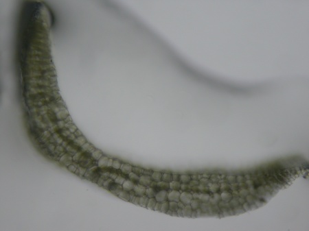
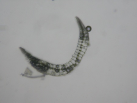
I’ve been meaning to have a close look at Leucobryum for a while, and am reminded of my intentions every day on the morning dog walk as I march past several good clumps of it on the banks and stumps at the south end of Kidbrooke Park. So this morning I collected some and had a good look.
When I’ve been out with Tom he’s often had a good sense of which Leucobryum species we have been looking at in the field, though I don’t have enough experience yet to tell them apart. However, I had at least picked up the sense that the relative lengths of the lower and upper parts of the leaf is not a reliable guide.
That was certainly true with my sample since the lower, broader part of the leaf was a bit shorter than the upper part. However, The Moss Flora of Britain and Ireland came to the rescue, noting that the costa in the lower part of the leaf is typically 4–5 cells thick in L. glaucum, and only 2 cells thick in L. juniperoideum. However, the paper in Journal of Bryology discussing the latter species in Britain notes that
care must be taken about exactly where the section is cut, for at higher levels there are only two layers of hyaline cells in both species.[1]
[Edit: See also Tom’s comment below, which adds extra important detail]
So, after a bit of fiddling around I managed to get a reasonable section, and confirmed it as L. glaucum (as I assumed).
Then, remembering that Tom showed me some L. juniperoideum in Wilderness Wood last year, which was also fruiting, I had a look at that too so I will know the difference in future.
But, while reading around I also see that the relation between morphology and genetics is complex,[2] which cast a bit of a blow to my excitement at making some headway with these plants.
[1] A. C. Crundwell (1972) Leucobryum juniperoideum (Brid.) C. Müll. in Britain, Journal of Bryology, 7:1, 1-5, DOI: 10.1179/jbr.1972.7.1.1
[2] A. Vanderpoorten, S. Boles, A.J. Shaw (2003) Patterns of morphological variation in Leucobryum albidum, L. glaucum, and L. juniperoideum (Bryopsida), Systematic Botany, 28,651-656, doi: http://dx.doi.org/10.1043/02-47.1. Available at: https://orbi.ulg.ac.be/bitstream/2268/168715/1/02-47.pdf


Nice photos Brad but possibly slightly misleading! It’s only the middle part of the leaf section you should be looking at. Thus, at its narrowest part in the middle, your L. glaucum section shows one cell on the ventral side of the ‘nerve’ and two cells on the dorsal side whereas the L. juniperoideum section shows just one cell either side of the central plane of chlorocysts. Away from the centreline of the leaf both species can exhibit marked thickening and many samples of L. juniperoideum look even more bulging (with hyalocysts) than your section of L. glaucum. These sections need to be cut about 1/4 to 1/3 of the way up the basal part of the leaf and can be tricky to interpret.
After spending many hours studying all published differences between these two species I eventually came to the conclusion that, in the absence of capsules, cell width is the most reliable single character and quite easy to use. You have to measure the width of the cells in the centre of the basal part of the leaf (i.e. half-way up the base) and you can do this without cutting a section – indeed, it’s best if you don’t. Measure the width of several adjacent hyalocyst cells on the ventral surface (the inside of the leaf) and take the average – so it’s just one actual measurement that’s needed. Take care not to over-flatten the leaf under the coverslip and don’t go right to the edge of the leaf where the cells are much narrower. Then you will find that L. juniperoideum has a mean cell width of 20 – 30 microns whereas L. glaucum measures 30 – 40 microns. It’s not completely foolproof and plants found on vertical sandrock are particularly troublesome. It works very well on stumps however where both species fruit more often than many bryologists give them credit for. There are numerous other differences, some of which you describe, and it’s prudent to use these too for additional confirmation, particularly when the cell width method gives a possibly ambiguous result (i.e. getting near 30 microns).
As many will know, Jacky Langton is studying this very subject for her M.Sc. and the results should be most interesting.
Tom
LikeLike
Thanks Tom! That is fantastically useful. I will try and revisit those samples again tomorrow.
LikeLike