
INTRODUCTION
An infectious disease is one of the serious diseases causing high mortality worldwide. For example, lower respiratory infections and diarrheal diseases remain the world’s most deadly infectious diseases, ranked as the fourth and eighth leading causes of death, respectively (World Health Organization, 2020). Commonly, microbial infections are treated with antibiotics. However, unfortunately, this has been increasingly complicated because some pathogenic bacteria having become more resistant to several antibiotics (Inggraini et al., 2021). Therefore, the finding of a new antimicrobial agent is a highly important subject to study.
Plants have been long used in the traditional treatment of microbial infections. Paederia foetida (Indonesian name: Sembukan; English name: King’s Tonic), a climbing herb from the Rubiaceae family, is one of Asia’s native medicinal plants found in both temperate and tropical Asia. The leaf of this plant has a bitter taste and is foul-smelling. Its leaf was traditionally used for the treatment of diarrhea, rheumatism, inflammation, piles, dysentery, and stomachache (Soni et al., 2013). Traditional knowledge provides great information for drug discovery.
The phytochemical and pharmacological activities of P. foetida have been investigated in earlier studies. Steroid, alkaloid, saponin, flavonoid, phenolic acids, tannin, paederolone, paederone, β-sitosterol, paederoside, glucosides, iridoid flavonoids, and volatile oils are the major substances found in its leaves (Mazumder et al., 2018; Patel, 2017). These compounds may contribute to the pharmacological activities of this plant. In addition, several biological activities of an extract derived from this plant have been studied at both in vitro and in vivo levels. In in vitro studies, the leaves extract of this plant was effective in inhibiting Escherichia coli (Silaban, 2021) and Vibrio cholerae (Hidayat et al., 2020). Several P. foetida fractions also showed antimicrobial activity against both Gram-positive and Gram-negative bacteria, as well as fungi (Morshed et al., 2012). In an in vivo study, this plant has been known to have a remarkable antihyperglycemic, antihyperlipidemic, and antioxidant activities in Wistar strain rats (Kumar et al., 2014). In addition, its leaf extract also was effective in inhibiting the infection of Aeromonas hydrophila on tilapia (Wahjuningrum et al., 2016). Although many reports were published about the pharmacological properties of P. foetida, particularly about its antibacterial activity, none of them identified the active compounds responsible for the antibacterial activity as well as their minimum inhibitory concentration (MIC) and minimum bactericidal concentration (MBC) values. In the present study, we evaluated the antibacterial and antibiofilm activities of the P. foetida methanolic leaves extract against several Gram-positive and Gram-negative bacteria. MIC and MBC determination, as well as identification of the most active fraction, was conducted to comprehensively investigate the antibacterial property of the crude extract and fractionated constituents.
MATERIALS AND METHODS
Plant material and bacterial strains
Fresh leaves of P. foetida Linn. were collected from Tasikmalaya, West Java, Indonesia (7°37?34.1?S 108°18?15.8?E). In addition, bacterial strains such as Bacillus subtilis DSM10, Staphylococcus aureus Newman, Mycobacterium smegmatis ATCC700084, E. coli BW25113, Pseudomonas aeruginosa PA14, and Acinetobacter baumannii DSM30008 were obtained from the Research Center for Pharmaceutical Ingredients and Traditional Medicine, National Research and Innovation Agency (BRIN), Kawasan PUSPIPTEK, Serpong, South Tangerang, Banten, Indonesia.
Preparation of crude extract
The collected leaves of P. foetida were gently washed with tap water. Then, the dried leaves were ground, and approximately 10 kg leaves were soaked in 100 ml methanol (1:10 w/v) for 24 hours at 28.5°C and agitated at 120 rpm. The mixture was then filtered and concentrated with a rotary evaporator at 40°C. The dry extract was then stored at 4°C before being used. For further analysis, it was dissolved in dimethyl sulfoxide (DMSO).
Antibacterial assay
Preliminary antibacterial activity screening was performed using the disk diffusion assay. Briefly, a suspension of bacterial inoculum was adjusted to McFarland standard 0.5 (equivalent to 1 × 10⁸ CFU/ml) and inoculated to a Mueller-Hinton agar (MHA, HiMedia) plate medium with sterile cotton buds. Then, the inoculated plates were allowed to dry at room temperature (±28°C) for ±10 minutes. Subsequently, sterile filter paper disks approximately 6 mm in diameter were impregnated with 10 µl of the crude extract (2 mg/ml) and placed on the surface of the agar plate. After incubation for 24 hours at 37°C, the antibacterial activity was evaluated by measuring the diameter of the zones of inhibition for microbial growth surrounding the disks. Tetracycline (0.1 mg/ml) served as the positive control. This assay was performed in triplicate.
Thin-layer chromatography (TLC) bioautography analysis
For TLC analysis, we used TLC silica gel 60 F254 precoated aluminum-backed TLC plates (10 cm × 2 cm with 0.2 mm thickness, Merck, Germany) as the stationary phase. Approximately 10 μl of the methanolic extract (0.032 g/ml) was spotted on the TLC plates. The TLC plates were developed containing a mixture of ethyl acetate and hexane (7:3 v/v) as the mobile phase. For TLC bioautography analysis, the extract was directly deposited (as bands) onto a TLC plate with total volume, concentration, and presaturated solvent as described earlier. The developed TLC plate was then removed from the solvents and dried at room temperature until dry. The TLC plate was then observed under UV light and 254 nm, and the dominant band was cut out into small pieces. Furthermore, the pieces of the TLC plates were then immersed in the plate MHA medium containing the bacterial culture and incubated for 24 hours at 37°C. Moreover, the active bands attributed to antibacterial activity were isolated using Preparative TLC Silica Gel GF, 500 microns, 20 × 20 cm (UNIPLATE Miles Scientific, USA). The active bands observed under UV light and 254 nm were scraped, collected, and purified by methanol.
Determination of MIC and MBC
The MIC value of the crude extract and active fractions was determined using a standard microdilution assay with some modifications (NCCLS, 2020). In short, two-fold dilutions of samples were supplemented to the 96-well sterile microtiter plate. Subsequently, the bacterial cells were set up in sterile normal saline and adjusted to McFarland standard 0.5, which is equivalent to 1 × 10⁸ CFU/ml, and it was then applied to each well of the plate. This particular plate was further incubated at 37°C for 24 hours. The MIC and MBC values were determined to be the lowest concentration of the extract that could suppress the bacterial growth, observed by the clear medium of the well and the particular concentration with no bacterial growth observed on the plate medium, respectively. Tetracycline (Sigma-Aldrich) was used as the positive control.
Antibiofilm assay
The potency of the crude extracts and fractions to inhibit the biofilm formation was carried out on a 96-well sterile microtiter plate (NCCLS, 2020). Shortly, 100 µl of bacterial suspensions (0.5 McFarland) was mixed into the wells containing the extract with different concentrations (2 × MIC, 1 × MIC, ½ × MIC, and ¼ × MIC) and the mixture of a brain heart infusion (BHI) medium along with incubation at 37°C for 24 hours. Furthermore, the bacterial cells in the suspension were removed, and the particular plate was washed twice with phosphate buffer saline (PBS). The wells were then stained with 200 µl of crystal violet (Sigma) (0.1%) after being air-dried, followed by incubation at 37°C for 30 minutes. The plate was further washed again with PBS. The stained biofilms were then solubilized with 200 µl of DMSO (99%). The absorbance that reflected the biofilm formation was detected at 595 nm, and the percentage of biofilm formation was then determined. The untreated bacterial culture was used as the negative control. Data were represented as mean ± standard deviation (n = 3).
Biofilm eradication analysis
The ability of the extracts and fractions to eradicate the established biofilm was examined by using a methyl tetrazolium test (MTT) following the particular method with slight modifications (Wintachai et al., 2019). In brief, 200 µl of bacterial cell suspension (1 × 10⁸ CFU/ml) was seeded into the flat bottom of a 96-well plate, followed by incubation at 37°C for 5 days. For biofilm production, during this period, the medium was frequently changed daily with 200 µl of a fresh BHI medium supplemented with 0.25% glucose. Further, samples in range concentration of ½ to 4 × MIC diluted with supplemented media were added to the wells and then incubated at 37°C overnight. After an incubation period, the medium was removed and substituted with 10 µl of the MTT solution (5 mg/ml) (Roche) along with incubation at 37°C for 4 hours. Soon thereafter, the insoluble formazan crystals were diluted with 200 µl of DMSO (99%). The absorbance was detected at 595 nm using an enzyme-linked immunoassay (ELISA) microplate reader, and data were represented as mean ± standard deviation (n = 3).
LC-MS/MS analysis of active fractions
The selected fractions were identified using the liquid chromatography- tandem mass spectrometry (LC-MS/MS) Xevo G2-XS quadrupole time of flight (QToF) mass spectrometry instrument (Waters, USA) via an electron spray interface. Chromatographic separation conditions were performed using an ultra performance liquid chromatography/QTof MS analytical system (Waters). Separation was reached by stepwise gradients from 95% A (0.1% formic acid and distilled water) and 5% B (acetonitrile and 0.1% formic acid) to 5% A and 95% B for 16 minutes. The flow rate of the desolvation gas was carried out to 1,000 l/hour, for cone gas, it was set to 50 l/hour, and the source temperature was fixed to 120°C. Capillary voltage and cone voltage were set to 2.0 and 30 kV, respectively. Mass spectrometry was determined using electrospray ionization Xevo G2-S QTof (Waters) with QToF mass spectrometry in positive ion mode. Moreover, the accurate mass and composition of the precursor ions and fragment ions were calculated and identified using the UniFi software library merged in the instrument (Septama et al., 2022).
Statistical analysis
The data were presented as means ± standard deviation from triplicates. One-way analysis of variance was used to compare the mean values with 95% and 99% confidence levels. Further analysis was performed using Tukey’s test, and p values < 0.05 were considered statistically significant.
RESULTS
Antibacterial activity of P. foetida Linn. leaves extract
The crude extract of P. foetida leaves showed antibacterial activity against E. coli and M. smegmatis as indicated by clear zone formation with diameters of 26 ± 1.4 and 37.6 ± 0.41 mm, respectively (Fig. 1). According to this assay, M. smegmatis was more sensitive to this extract than E. coli. On the contrary, there is no antibacterial activity of extracts on other bacterial strains, including P. aeruginosa, A. baumannii, B. subtilis, and S. aureus (Table 1). As a positive control, tetracycline exhibited a broad spectrum of Antibacterial activity in all bacterial strains used in this study, with a clear zone diameter ranging from 28 ± 1.2 to 30 ± 1.6 mm.
Fractions of P. foetida-derived extract and their antibacterial activities
The chemical components in the crude extract of P. foetida have successfully been fractionated into six different fractions in TLC. These fractions were coded as F1, F2, F3, F4, F5, and F6. The separated spot and retention factor (Rf) values of each fraction are shown in Figure 2. The yield of these fractions was pasta. Each fraction was subjected to a bioautography antibacterial assay. The F5 and F6 fractions exhibited antibacterial activity against both E. coli and M. smegmatis (Fig. 3). These fractions were more active in M. smegmatis than in E. coli, as indicated by the broader clear zone formation in M. smegmatis. Meanwhile, antibacterial activity was not found in other fractions. Therefore, F5 and F6 were considered as active fractions.
 | Figure 1. Antibacterial activity of P. foetida leaves extract against (A) E. coli and (B) M. smegmatis. A1 and B4: tetracycline (0.1 mg/ml); A2, B1, B2, and B3: P. foetida leaves extract; and B5: distilled water. [Click here to view] |
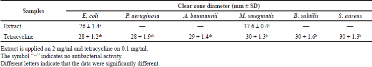 | Table 1. Antibacterial activity of P. foetida Linn. leaves extract. [Click here to view] |
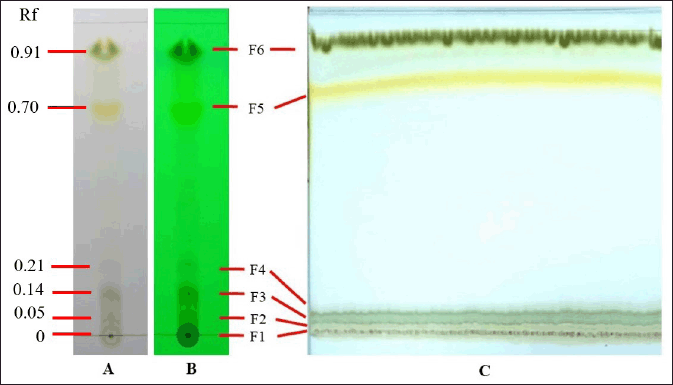 | Figure 2. TLC analysis (10 × 2 cm) of the methanol-derived P. foetida extract using a mobile phase eluent mixture of ethyl acetate and n-hexane (7:3 v/v). Ten microliters of the 0.032 g/ml extract diluted with methanol was applied as the straight line and visualized (A) under UV light and (B) under UV 254 nm. (C) Preparative TLC (20 × 20 cm) visualized under UV light; 3,500 μl of 0.042 g/ml of P. foetida extract was applied. Six fraction spots with different Rf values were separated. [Click here to view] |
MIC and MBC of crude extract and active fractions of P. foetida leaves
The crude extract and active fractions (F5 and F6) of P. foetida leaves have different MIC and MBC toward E. coli and M. smegmatis (Table 2). In general, the F5 and F6 fractions have lower MIC and MBC than those of the crude extract. The F6 fraction has the lowest MIC and MBC against two tested bacterial strains and is more active with M. smegmatis. The MIC and MBC of the F6 fraction against M. smegmatis were 23.43 and 31.25 µg/ml, respectively. To confirm this result, bacterial growth inhibition has also been shown by tetracycline.
Antibiofilm activity and eradication of bacterial biofilm growth cells from crude extract and active fractions of P. foetida leaves
The effects of the P. foetida leaves extract and active fractions on inhibition and eradication of biofilm are shown in Figure 4. The crude extract, F5 and F6 fractions, of P. foetida leaves showed diverse antibiofilm properties against both E. coli and M. smegmatis. The higher the extract or fraction concentrations applied, the lower the percentage of bacterial biofilm formation. All samples exhibited the best antibiofilm activity against two bacteria tested in two times of their MIC. In this concentration, more than ±50% of M. smegmatis biofilm and more than 30% of E. coli biofilm have been inhibited. Therefore, M. smegmatis was likely more sensitive than E. coli to all samples applied. Two times of the F5 fraction MIC showed the lowest percentages of biofilm formation both in M. smegmatis and in E. coli with percentages of biofilm formation of 39.7% and 65.1%, respectively. In addition, all samples also showed biofilm eradication activity against the tested strains. The lowest percentage of viability of bacterial biofilm-grown cells was found in two times of MIC. The two MIC of the F5 fraction was effective in the eradication of M. smegmatis biofilm, while F6 was effective in eradicating E. coli biofilm.
Chemical components in active fractions of P. foetida leaves
Several compounds were identified as major compounds in the most active fractions, F5 and F6. Linolenic acid, carotenoid, icosanamide, and two unknown compounds were detected in the F5 fraction, while phaeophytin A and three unknown compounds were detected in the F6 fraction. Those compounds have various retention times and abundance in each fraction as exhibited by the liquid chromatography-tandem mass spectrometry (LC-MS/MS) chromatogram (Fig. 5).
DISCUSSION
An antibacterial agent originating from natural sources is becoming an urgent need for treating infectious diseases caused by pathogenic bacteria. Medicinal plants could be a great source of new antibacterial compounds. This study evaluated the potency of P. foetida leaves-derived extract against some bacterial strains, such as E. coli, M. smegmatis, P. aeruginosa, A. baumannii, B. subtilis, and S. aureus. The crude extract of this plant showed promising antibacterial activity towards E. coli and M. smegmatis and no antibacterial activity against other bacteria, showing the selective actions of the extract. The effectivity of P. foetida leaves on all bacteria tested may be influenced by the resistance of each bacterial strain to the extract in that concentration. In this study, 2 mg/ml of the P. foetida methanolic extract was effective in inhibiting the growth of E. coli and M. smegmatis in vitro. Supporting these results, earlier studies also found that the ethanolic extract of P. foetida also could inhibit several bacteria, such as Erwinia carotovora, Xanthomonas campestris, and Ralstonia solanacearum, with the extract concentration of 100 mg/ml (Namsena et al., 2019), and Enterococcus faecalis, S. aureus, Shigella flexneri, and E. coli in the concentration of 25 mg/ml (Uddin et al., 2007). Therefore, both methanol and ethanol could be used as the solvent for extracting antibacterial compounds originating from this plant.
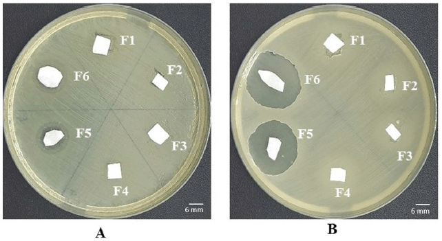 | Figure 3. Antibacterial activity of the selected fraction against (A) E. coli and (B) M. smegmatis. [Click here to view] |
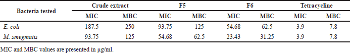 | Table 2. MIC and MBC values of extract and active fractions of P. foetida leaves. [Click here to view] |
To investigate the active components of the P. foetida leaves extract responsible for antibacterial action, six fractions with different retention factors have been separated in the TLC plate, namely, F1, F2, F3, F4, F5, and F6. Among these fractions, F5 and F6 were the most active fractions in inhibiting E. coli and M. smegmatis. Interestingly, these two fractions exhibited greater antibacterial activity against M. smegmatis than E. coli. These results indicate that M. smegmatis are more sensitive to these fractions. This finding is interesting because M. smegmatis have been resistant to some β-lactam antibiotics (Nguyen et al., 2017). Therefore, these fractions could be the alternative source of an anti-M. smegmatis agent in the future.
In the present study, a comprehensive investigation was carried out to evaluate the MIC and MBC of the P. foetida leaves extract and its active fractions to E. coli and M. smegmatis. In general, two fractions, F5 and F6, had lower MIC and MBC to the tested bacteria than the crude extract. This result likely suggests that purified compounds are more active than the crude extract. Consistently, the F5 and F6 fractions displayed the strongest antibacterial activity on M. smegmatis, as indicated by the lowest of their MIC and MBC on that particular bacterium. The MIC and MBC of these fractions to tested bacteria ranged from 23.43 to 125 µg/ml, whereas the MIC and MBC of the crude extract had higher concentrations ranging from 93.75 to 250 µg/ml. The MIC and MBC of the extract <5 mg/ml are considered as strong antibacterial activity (Bussmann et al., 2010).
Certain bacteria developed to be more resistant to available antibiotics through biofilm formation (Fuente-Núñez et al., 2013). Escherichia coli is well known as a biofilm former in urinary tract infections (Sharma et al., 2016), and M. smegmatis, a biofilm former, also has been widely studied as a surrogate microorganism used in antimycobacterial drug discovery assays (Bhunu et al., 2017). Fortunately, the crude extract and active fractions of P. foetida leaves possessed strong antibiofilm activity with dual mechanisms in preventing and eradicating the biofilm of E. coli and M. smegmatis. Among all samples tested, the F5 and F6 fractions were more effective in reducing the biofilm formation of the tested bacteria than the crude extract at the same concentration. Similarly, it has been reported that the methanolic extract from other plant sources, such as Bergenia ciliata, Prosopis laevigata, Opuntia ficus-indica, and Gutierrezia microcephala, also can reduce bacterial biofilm (Alam et al., 2020; Sánchez et al., 2016). Moreover, the extract and fractions derived from P. foetida leaves were also able to destroy the biofilm mass of the tested bacteria. The two MIC of the F5 fraction was effective in eradicating M. smegmatis biofilm, while F6 was effective in eradicating E. coli biofilm. These results indicate the selective action of each isolated fraction. However, both preventing and destroying biofilm are important mechanisms for reducing the virulence factor of certain pathogenic bacteria. Therefore, the crude extract and active fractions of this plant possibly provide new candidates for antimicrobial agents in the future.
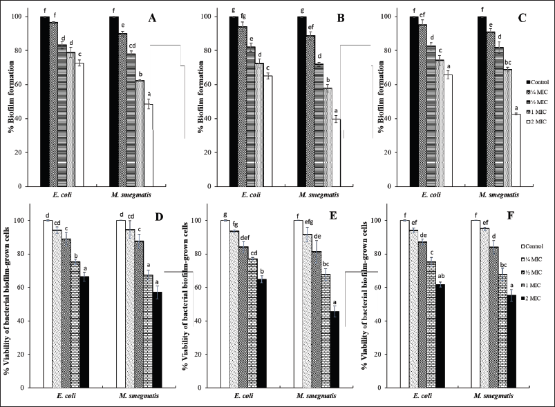 | Figure 4. Antibiofilm activity of (A) crude extract, (B) F5, and (C) F6; biofilm eradication activity of (D) extract, (E) F5, and (F) F6. [Click here to view] |
 | Figure 5. LC-MS/MS chromatogram of active fractions (A) F5 and (B) F6 derived from P. foetida. [Click here to view] |
To get a deep understanding of the chemical composition of the most active fractions of the P. foetida leaves extract, LCMS/MS analysis has been carried out. Two fractions, F5 and F6, have different major components which are well investigated as antibacterial compounds. The F5 fraction was dominated by linolenic acid, carotenoid, and icosanamide. An earlier study reported linolenic acid displayed antibacterial activity against B. subtilis and S. aureus (Kusumah et al., 2020). Carotenoids extracted from Rhodotorula glutinis have an antibacterial effect against S. aureus, B. subtilis, B. cereus, Salmonella enteritidis, and E. coli (Keceli et al., 2013). However, there is no comprehensive report on the biological activity of icosanamide. In addition, the F6 fraction was dominated by phaeophytin A. This compound also has been well studied to have antimicrobial action against S. aureus, E. coli, Streptococcus pneumoniae, Salmonella typhi, and Candida albicans (Ekalu et al., 2019). However, both the F5 and F6 fractions contained unknown compounds, which are possibly new compounds.
CONCLUSION
This study concluded that the P. foetida leaves extract and its fractions (F5 and F6) have antibacterial activity against E. coli and M. smegmatis. The F5 and F6 fractions possessed stronger antibacterial activity than the crude extract. Besides, these two active fractions also have stronger antibiofilm activity by preventing biofilm formation and eradicating the established biofilm. All these fractions are contained by active compounds known as antibacterial agents. Further study is necessary to purify and develop the antibacterial property of these active fractions in terms of providing new antibacterial agents.
ACKNOWLEDGMENTS
All authors thank the Division of Microbiology, Department of Biology, Faculty of Mathematics and Natural Sciences, IPB University, and the National Research and Innovation Agency (BRIN), Indonesia, as well as the Ministry of Education and Culture, Republic of Indonesia, through Student Creativity Program 2021, for supporting this study.
CONFLICTS OF INTEREST
All authors declare there are no conflicts of interest related to this study.
FUNDING
This study was partly funded by The Ministry of Education and Culture Indonesia through Student Creativity Program 2021, and Partly funded by National Priority Program, Deputy for Engineering Science, Indonesian Institute of Sciences (LIPI), Indonesia, 2021 [Grant Number: 58/A/DT/2021].
AUTHORS’ CONTRIBUTIONS
JAP, MEP, GSS conceived of presented idea and developed the experimental design. JAP, GSS, WD, TYA, AIAY, EA contributed to sample preparation. JAP, MEP, GSS, WD, TYA, AIAY, EA, ZPT carried out laboratory work and data analysis. JAP, MEP, GSS, WD, TYA, AIA, EA verified and interpreted all data. JAP, MEP, ZPT, and TM contributed in paper writing.
ETHICAL APPROVALS
This study does not involve experiments on animals or human subjects.
DATA AVAILABILITY
All data generated and analyzed are included within this research article.
PUBLISHER’S NOTE
This journal remains neutral with regard to jurisdictional claims in published institutional affiliation.
REFERENCES
Alam K, Al Farraj DA, Mah-E-Fatima S, Yameen MA, Elshikh MS, Alkufeidy RM, Mustafa AEZMA, Bhasme P, Alshammari MK, Alkubaisi NA, Abbasi AM, Naqvi TA. Anti-biofilm activity of plant derived extracts against infectious pathogen-Pseudomonas aeruginosa PAO1. J Infect Publ Health, 2020; 13(11):1734–41; doi:10.1016/j.jiph.2020.07.007. CrossRef
Bhunu B, Mautsa R, Mukanganyama S. Inhibition of biofilm formation in Mycobacterium smegmatis by Parinari curatellifolia leaf extracts. BMC Complement Altern Med, 2017; 17(285):1–10; doi:10.1186/s12906-017-1801-5. CrossRef
Bussmann RW, Malca-García G, Glenn A, Sharon D, Chait G, Díaz D, Pourmand K, Jonat B, Somogy S, Guardado G, Aguirre C, Chan R, Meyer K, Kuhlman A, Townesmith A, Effio-Carbajal J, Frías-Fernandez F, Benito M. Minimum inhibitory concentrations of medicinal plants used in Northern Peru as antibacterial remedies. J Ethnopharmacol, 2010; 132(1):101–8; doi:10.1016/j.jep.2010.07.048. CrossRef
Ekalu A, Ayo RGO, Habila JD, Hamisu I. Bioactivities of phaeophytin a, α-amyrin, and lupeol from Brachystelma togoense Schltr. J Turk Chem Soc, 2019; 6(3):411–8. CrossRef
Fuente-Núñez CDL, Reffuveille F, Fernández L, Hancock REW. Bacterial biofilm development as a multicellular adaptation: antibiotic resistance and new therapeutic strategies. Curr Opin Microbiol, 2013; 16(5):580–9; doi:10.1016/j.mib.2013.06.013. CrossRef
Hidayat AF, Duniaji AS, Arihantana NMIH. Uji daya hambat ekstrak daun sembukan (Paederia foetida) terhadap Vibrio cholerae [Indonesian]. J Itepa, 2020; 9(4):390–9. CrossRef
Inggraini M, Nurfajriah S, Priyanto JA, Ilsan NA. Antimicrobial susceptibility and molecular species identification of clinical carbapenem-resistant bacteria. Biodivers J Biol Divers, 2021; 22(2):555–62. CrossRef
Keceli TM, Erginkaya Z, Turkkan E, Kaya U. Antioxidant and antibacterial effects of carotenoids extracted from Rhodotorula glutinis strains. Asian J Chem, 2013; 25(1):42–6; doi:10.14233/ajchem.2013.12377. CrossRef
Kumar V, Anwar F, Ahmed D, Verma A, Ahmed A, Dmanhouri ZA, Mishra V, Wamteke PW, Bhatt PC, Mujeeb M. Paederia foetida Linn. leaf extract: an antihyperlipidemic, antihyperglycaemic and antioxidant activity. BMC Complement Altern Med, 2014; 14(76):1–16. CrossRef
Kusumah D, Wakui M, Murakami M, Xie X, Yukihito K, Maeda I. Linoleic acid, α-linolenic acid, and monolinolenins as antibacterial substances in the heat-processed soybean fermented with Rhizopus oligosporus. Biosci Biotech Biochem, 2020; 84(6):1285–90; doi:10.1080/09168451.2020.1731299.Mazumder K, Dey ZK, Dey S, Hossain MDS, Sajon SR, Hossain KMD. Phytochemical screening & evaluation of ant diarrheal mode of action of leaves extracts of Paederia foetida Linn. Int J Complement Altern Med, 2018; 11(5):304–9; doi:10.15406/ijcam.2018.11.00416. CrossRef
Morshed H, Isla MS, Parvin S, Ahmed MU, Islam MS, Mostofa AGM, Sayeed MSB. Antimicrobial and cytotoxic activity of the methanol extract of Paederia foetida Linn. (Rubiaceae). J Appl Pharmaceut Sci, 2012; 2(1):77–80.
Namsena P, Maneetap J, Phonpheng D, Ngamsane W. Comparative study of antibacterial activity and phytochemical analysis of stem, root and leaf extracts of Paederia foetida L. against phytopathogenic bacteria. Agr Nat Resour, 2019; 53:395–401.
NCCLS (National Committee for Clinical Laboratory Standard). Performance standard for antimicrobial susceptibility testing. Ninth informational supplement. 30th edition, NCCLS, Malvern, PA, vol. 40, no. 1, pp 1–294, 2020.
Nguyen TV, Blackledge MS, Lindsey EA, Minrovic BM, Ackart DF, Jenon AB, Obregón-Henao A, Melander RJ, Basaraba RJ, Melander C. The discovery of 2-aminobenzimidazoles that sensitize Mycobacterium smegmatis and M. tuberculosis to β-lactam antibiotics in a pattern distinct from β-lactamase inhibitors. Angew Chem Int Ed Engl, 2017; 56(14):3940–4; doi:10.1002/anie.201612006. CrossRef
Patel DK. Paederia Foetida Linn: a potential climbing medicinal herb in Central India. Int J Environ Sci Nat Resour, 2017; 6(5):118–24.
Sánchez E, Morales CR, Castillo S, Leos-Rivas C, García-Becerra L, Martínez DMO. Antibacterial and antibiofilm activity of methanolic plant extracts against nosocomial microorganisms. Evid Based Complement Altern Med, 2016; 2016:1–8; doi:10.1155/2016/1572697. CrossRef
Septama AW, Tasfiyati AN, Kristiana R, Jaisi A. Chemical profiles of essential oil from Javanese turmeric (Curcuma xanthorrhiza Roxb.). Evaluation of its antibacterial and antibiofilm activities against selected clinical isolates. South Afr J Bot, 2022; 146:728–34; doi:10.1016/j.sajb.2021.12.017. CrossRef
Sharma G, Sharma S, Sharma P, Chandola D, Dang S, Gupta S, Gabrani R. Escherichia coli biofilm: development and therapeutic strategies. J Appl Microbiol, 2016; 121(2):309–19; doi:10.1111/jam.13078. CrossRef
Silaban H. The effect of various concentrations of ethanol extract of the leaves of Paederia foetida L. on the growth of Escherichia coli bacteria. J Drug Deliv Ther, 2021; 11(6):61–7. CrossRef
Soni RK, Irchaiya R, Dixit V, Alok S. Paederia foetida Linn: phytochemistry, pharmacological and traditional uses. Int J Pharmaceut Sci Res, 2013; 4(12):4525–30.
Uddin B, Nahar T, Khalil MI, Hossain S. In vitro antibacterial activity of the ethanol extract of Paederia foetida L. (Rubiaceae) leaves. Bangladesh J Life Sci, 2007; 19(2):141–3.
Wahjuningrum D, Hasanah M, Rahman. Effication of skunkvine leaves Paederia foetida for prevention of Aeromonas hydrophila infection on tilapia. J Akuakult Indones, 2016; 15(2):108–16. CrossRef
World Health Organization. The top 10 causes of death. 2020. [ONLINE] Available via https://www.who.int/news-room/fact-sheets/detail/the-top-10-causes-of-death (Accessed 21 December 2021).
Wintachai P, Paosen S, Yupanqui CT, Voravuthikunchai SP. Silver nanoparticles synthesized with Eucalyptus critriodora ethanol leaf extract stimulate antibacterial activity against clinically multidrug-resistant Acinetobacter baumannii isolated from pneumonia patients. Microb Pathog, 2019; 126:245–57. CrossRef