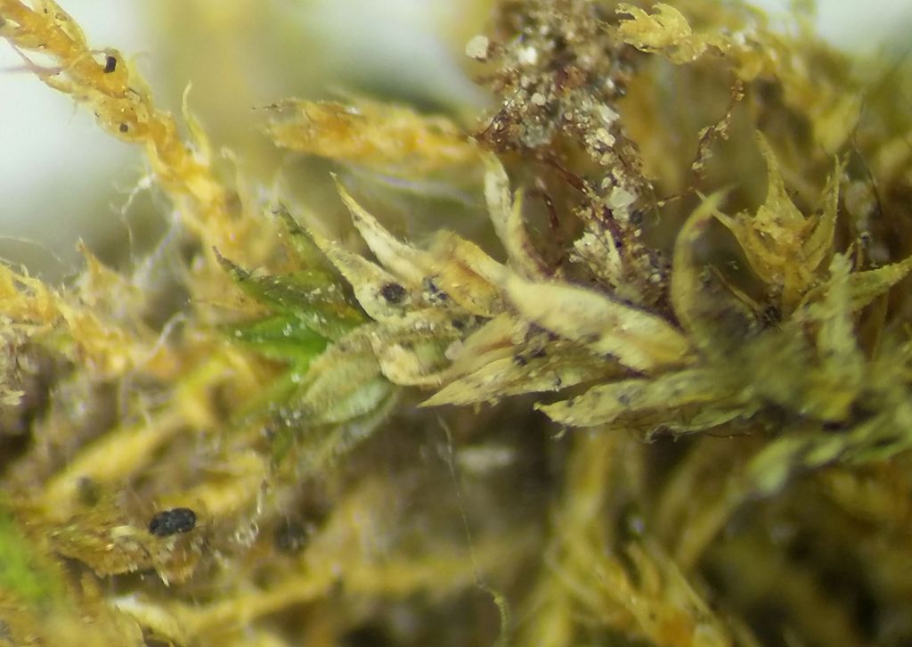
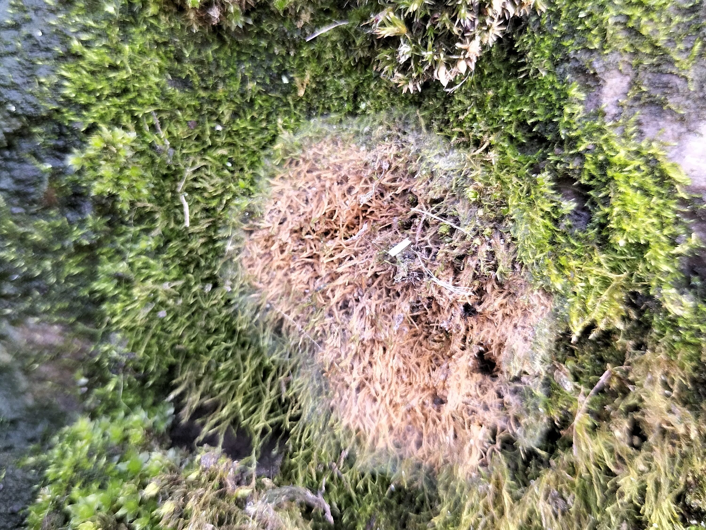
Host bryophytes: Amblystegium serpens, Tortula muralis and Didymodon cf rigidulus [England], along with Platygyrium repens, Brachythecium sp., Pylaisia polyantha and Anomodon rugelii in mainland Europe. However, the English collection was strongly associated with the bryophilous fungus Acrospermum adeanum, raising the possibility that this fungus may be mycoparasitic on that rather than the mosses.
My draft description, also featured on a post on AscoFrance:
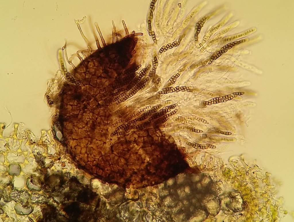
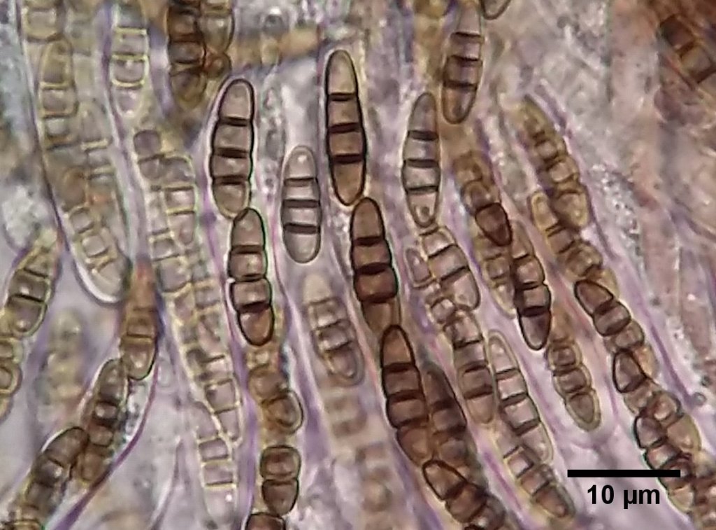
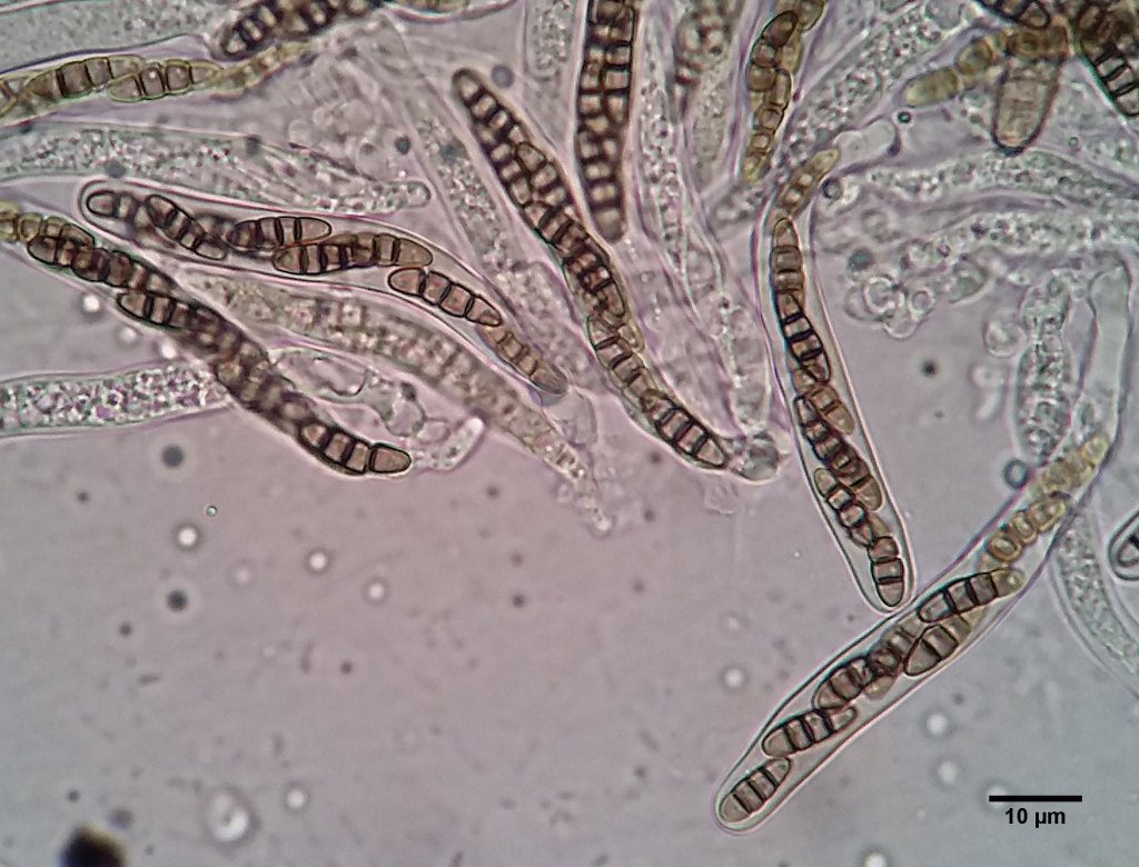
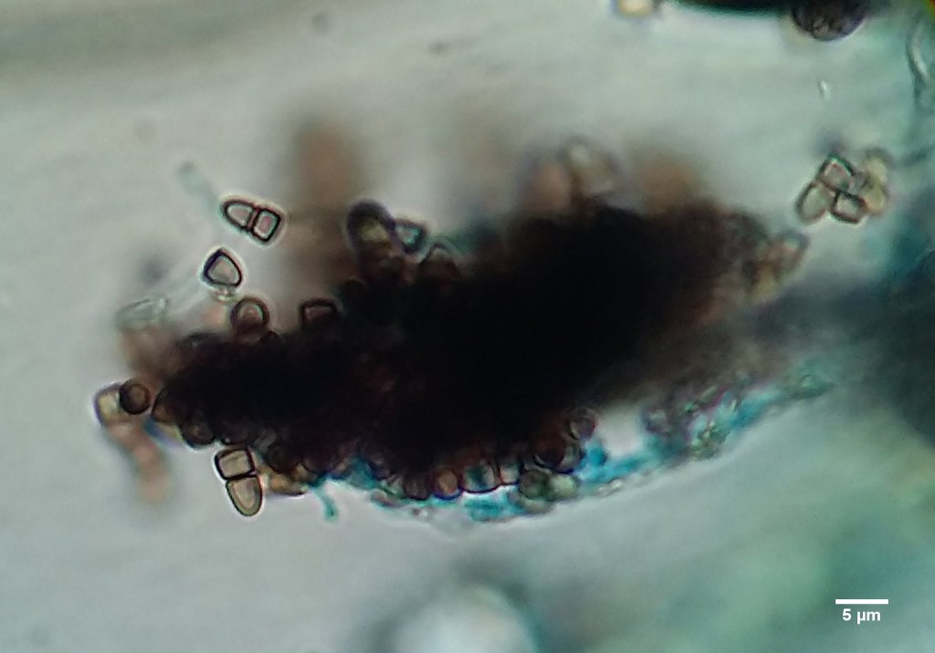
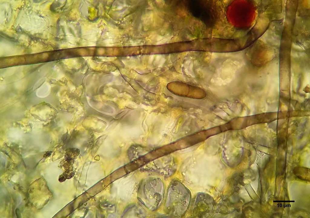
Teleomorph: Ascomata up to ~100um diam, setose, solitary. Hairs brown below, occasionally branched below, paler above, walls verrucose, length seems variable, up to and in excess of 40um. Peridium outer cells textura angularis. Asci 8-spored, clavate-cylindrical, around 110 x 10, bitunicate. Mature ascospores brown, 3-septate, fragmenting in asci and after dispersal = pseudopolysporous. Spore length and width increase somewhat during maturation, apparently along with the strength of the constrictions at the septa. Spores 11.5-13.6 x 3.4-3.9.
Anamorph: Conidiophores seem to be unbranched with verrucose walls. Some may form on top of the fruitbody from older hairs but I am not sure. Others were definitely beside the fruitbodies on the substrate. I think they were >100um long, 5 wide. Forming conidia apically in short chains but possibly also laterally. Conidia brown, verrucose, (0-)1-septate, 14.0-19.2 x 5.1-6.5(-7.2).
I am uncertain about whether the anamorph is the same fungus as it has not been described previously, but will currently treat it so due to the strong similarities in colour, wall ornamentation and co-localisation.